Call us
301-363-4651 (Available 9 a.m. to 5 p.m. CST from Monday to Friday)
PSMA1 encodes the proteasome subunit alpha type-1 protein. Also known as Proteasome alpha 1 subunit, it is a crucial component of the 20S core proteasome. This protein plays a key role in the proteolytic machinery of cells by facilitating the degradation of damaged or misfolded proteins. It participates in the ubiquitin - proteasome pathway, which is essential for maintaining cellular homeostasis.
Alterations in PSMA1 have been associated with various diseases, including neurodegenerative disorders and certain cancers. In drug development, targeting the proteasome complex, including PSMA1, has become an area of interest. Some proteasome inhibitors have shown potential in pre - clinical and clinical trials for treating cancer, aiming to disrupt the abnormal protein degradation in tumor cells.
PSMA1 Recombinant Monoclonal Antibody (CSB-RA276081A0HU)
Validated Data

IHC image of CSB-RA276081A0HU diluted at 1:100 and staining in paraffin-embedded human colorectal cancer performed on a Leica BondTM system. After dewaxing and hydration, antigen retrieval was mediated by high pressure in a citrate buffer (pH 6.0). Section was blocked with 10% normal goat serum 30min at RT. Then primary antibody (1% BSA) was incubated at 4°C overnight. The primary is detected by a Goat anti-rabbit polymer IgG labeled by HRP and visualized using 0.05% DAB.

IHC image of CSB-RA276081A0HU diluted at 1:100 and staining in paraffin-embedded human small intestine tissue performed on a Leica BondTM system. After dewaxing and hydration, antigen retrieval was mediated by high pressure in a citrate buffer (pH 6.0). Section was blocked with 10% normal goat serum 30min at RT. Then primary antibody (1% BSA) was incubated at 4°C overnight. The primary is detected by a Goat anti-rabbit polymer IgG labeled by HRP and visualized using 0.05% DAB.
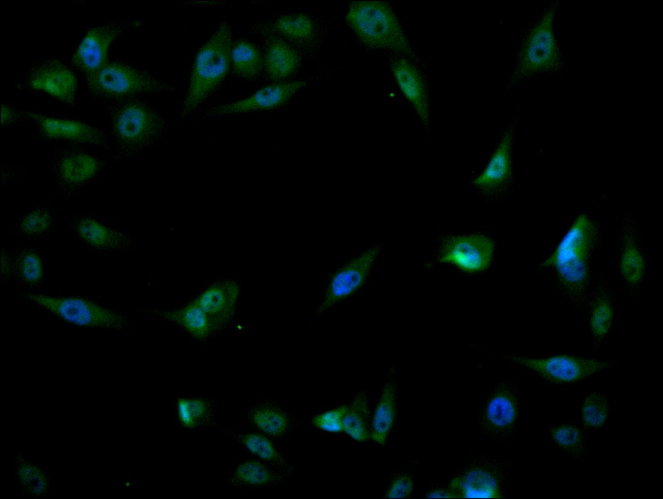
Immunofluorescence staining of PC-3 cell with CSB-RA276081A0HU at 1:50, counter-stained with DAPI. The cells were fixed in 4% formaldehyde, permeabilized using 0.2% Triton X-100 and blocked in 10% normal Goat Serum. The cells were then incubated with the antibody overnight at 4°C. The secondary antibody was Alexa Fluor 488-congugated AffiniPure Goat Anti-Rabbit IgG(H+L).
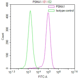
Overlay Peak curve showing HepG2 cells stained with CSB-RA276081A0HU (red line) at 1:100. The cells were fixed in 4% formaldehyde and permeated by 0.2% TritonX-100 for 10min. Then 10% normal goat serum to block non-specific protein-protein interactions followed by the antibody (1ug/1*106cells) for 45min at 4℃. The secondary antibody used was FITC-conjugated goat anti-rabbit IgG (H+L) at 1/200 dilution for 35min at 4℃.Control antibody (green line) was Rabbit IgG (1ug/1*106cells) used under the same conditions. Acquisition of >10,000 events was performed.
The following PSMA1 reagents supplied by CUSABIO are manufactured under a strict quality control system. Multiple applications have been validated and solid technical support is offered.
PSMA1 Antibodies for Homo sapiens (Human)
| Code | Product Name | Species Reactivity | Application |
|---|---|---|---|
| CSB-PA018865GA01HU | PSMA1 Antibody | Human,Mouse,Rat | ELISA,WB,IHC |
| CSB-PA018865LA01HU | PSMA1 Antibody | Human | ELISA, WB, IHC, IF, IP |
| CSB-PA018865LB01HU | PSMA1 Antibody, HRP conjugated | Human | ELISA |
| CSB-PA018865LC01HU | PSMA1 Antibody, FITC conjugated | Human | |
| CSB-PA018865LD01HU | PSMA1 Antibody, Biotin conjugated | Human | ELISA |
| CSB-RA276081A0HU | PSMA1 Recombinant Monoclonal Antibody | Human | ELISA, IHC, IF, FC |
PSMA1 Proteins for Homo sapiens (Human)
| Code | Product Name | Source |
|---|---|---|
| CSB-YP018865HU CSB-BP018865HU CSB-MP018865HU CSB-EP018865HU-B |
Recombinant Human Proteasome subunit alpha type-1 (PSMA1) | Yeast Baculovirus Mammalian cell In Vivo Biotinylation in E.coli |
| CSB-EP018865HU | Recombinant Human Proteasome subunit alpha type-1 (PSMA1), partial | E.coli |

