Call us
301-363-4651 (Available 9 a.m. to 5 p.m. CST from Monday to Friday)
| Code | CSB-RA902876A0HU |
| Size | US$210 |
| Order now | |
| Image |
|
| Have Questions? | Leave a Message or Start an on-line Chat |
| Application | Recommended Dilution |
|---|---|
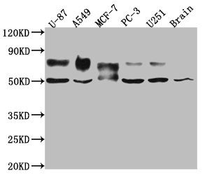
| WB | 1:500-1:5000 |
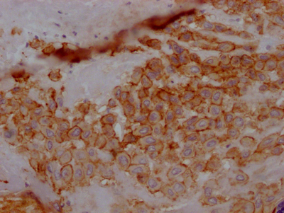
| IHC | 1:50-1:200 |
The TPBG recombinant monoclonal antibody is a highly specific anti-TPBG antibody. This TPBG antibody was produced by inserting the DNA sequence encoding the TPBG monoclonal antibody into plasmids and then transfecting the plasmids into cell lines for expression. Its isotype is identical to rabbit IgG. This TPBG antibody detects the TPBG protein in human and mouse samples and can be used in ELISA, WB, and IHC.
TPBG, an oncofetal antigen, is low expressed in normal tissues but upregulated in a variety of human cancers. High expression of TPBG usually indicates a poor clinical outcome. Jeong-Eun Yoo et. al has found that TPBG is an efficient sorting marker for ventral mesencephalic dopaminergic precursors originated from human pluripotent stem cells.
There are currently no reviews for this product.

