Call us
301-363-4651 (Available 9 a.m. to 5 p.m. CST from Monday to Friday)
The Enzyme-Linked Immunosorbent Assay (ELISA) is a foundational immunochemical technique renowned for its high specificity, sensitivity, and relative operational simplicity. Its core principle relies on the specific binding between an antigen and its antibody, with detection achieved via an enzyme conjugated to an antibody that catalyzes a colorimetric or chemiluminescent signal upon substrate addition, enabling qualitative and quantitative analysis of target molecules [1,2]. As a pivotal tool, ELISA plays a central role across diverse fields, including basic scientific research, clinical diagnostics, environmental monitoring, and drug development.
While numerous ELISA variants exist, they are fundamentally derived from four classic types: Direct, Indirect, Sandwich, and Competitive ELISA [1,2]. A deep understanding of these formats is crucial for selecting the appropriate experimental protocol, accurately interpreting results, and leveraging the full potential of this versatile platform.
Table of Contents
2. Innovations and Frontiers in ELISA Technology
3. Evolving Applications of ELISA: From Lab Bench to Real World
Through constant optimization, several ELISA formats have been developed to cater to different applications, primarily distinguished by their detection principles and procedural steps. The four fundamental types are direct, indirect, Sandwich, and competitive ELISA.
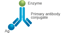
In direct ELISA, only an enzyme-labeled primary antibody is used. The enzyme-conjugated primary antibody "directly" binds to the antigen in the sample that is immobilized to the plate. Next, the enzyme-labeled primary antibody reacts with its substrate to produce a visible, detectable signal, the intensity of which is directly proportional to the amount of antigen present in the sample [2].
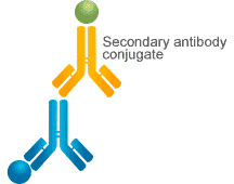
In indirect ELISA, both an unlabeled primary antibody and an enzyme-conjugated secondary antibody are used.
The primary antibody binds to the antigen immobilized to the plate, and then the enzyme-conjugated secondary antibody binds to the primary antibody. Finally, the enzyme linked to the secondary antibody reacts with its substrate to produce a visible signal that can be measured.
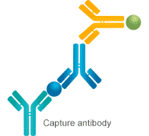
The sandwich ELISA employs two antibodies that bind to distinct, non-overlapping epitopes on the target antigen. The "capture antibody" is pre-coated onto the plate to immobilize the antigen from the sample. In indirect Sandwich ELISA, the unlabeled primary detection antibody is added and binds to the captured antigen, forming an antibody-antigen-antibody "sandwich". The enzyme-conjugated secondary antibody is then introduced, and a visible signal is generated upon substrate addition [2]. The signal intensity correlates directly with the amount of captured antigen.
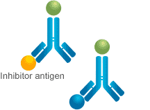
The competitive ELISA, also known as inhibition ELISA, involves the target antigen competing with plate-immobilized reference antigen for binding to a limited amount of enzyme-labeled antibody. The amount of labeled antibody that binds to the plate is inversely proportional to the concentration of antigen in the sample. Therefore, a higher sample antigen concentration results in less antibody binding and a weaker measured signal upon substrate addition [2].
There is another type of competitive ELISA based on antigen capture, in which the plate is coated with an unlabeled antibody. In this case, a known quantity of enzyme-labeled antigen and antigen in the sample compete for binding to pre-coated antibody.
Table 1. Advantages and disadvantages of each ELISA type
|
|
Advantages | Disadvantages |
|---|---|---|
| Direct ELISA |
|
|
| Indirect ELISA |
|
|
| Sandwich ELISA |
|
|
| Competitive ELISA |
|
|
Choosing the optimal ELISA format depends on several key factors: the nature of the analyte (size, availability, purity), the required assay performance (sensitivity, specificity), the available reagents (antibodies), and the experimental context (throughput, cost). The following logic flow provides a practical guide for selection:
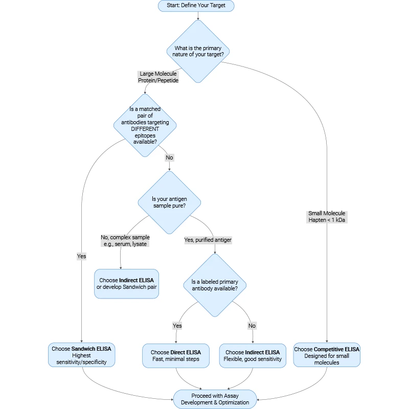
Figure: Selection Decision Guide of Four ELISA types
Choosing the optimal ELISA format depends on several key factors: the nature of the analyte (size, availability, purity), the required assay performance (sensitivity, specificity), the available reagents (antibodies), and the experimental context (throughput, cost). The following logic flow provides a practical guide for selection:
Recent ELISA innovations after 2020 have centered on re‑engineering assay architectures, embedding powerful biochemical amplification modules, and migrating from simple colorimetric plate readers to multiplexed optical, electrochemical, and AI‑assisted detection systems.
Recent work has shifted ELISA away from static 96‑well plates toward integrated microfluidic and digital‑microfluidic (DMF) formats that automate multistep workflows, minimize sample volume, and support high‑throughput multiplexing [4,5].
Beyond chip‑scale fluidics, “intelligent” device design is emerging, combining microfluidics with embedded computation and AI‑driven control.
In parallel, there is a sustained effort in surface and capture‑chemistry design within traditional plate formats [5].
Modern ELISA development increasingly treats the immunocomplex as a trigger for programmable biochemical amplification rather than a direct link to a single enzyme reaction [5,6].
Nanomaterial‑based and electrochemiluminescent (ECL) amplification strategies have also advanced rapidly [5].
Another frontier is "in situ amplification" of protein targets themselves rather than solely amplifying reporter signals [6].
Synthetic‑biology concepts increasingly intersect with CRISPR‑based detection [5,7].
Detection has evolved from simple absorbance measurements to a diverse landscape of optical, electrochemical, and digital readouts designed for higher sensitivity, multiplexing, and automation [4,5].
Colorimetric ELISA remains dominant in many settings, but next‑generation platforms are rapidly adopting fluorescence and chemiluminescence to extend dynamic range and lower detection limits.
Time‑domain and dynamic‑range engineering is another active area.
Digital ELISA (dELISA) converts analog enzyme activity into binary events in tiny volumes so that individual molecules can be counted [5,9].
Digital imaging and analysis are increasingly paired with machine learning.
Electrochemical readouts form a parallel frontier, particularly within microfluidic ELISA [5].
Hybrid systems also emerge where ELISA‑style capture interfaces with high‑information‑content detectors [9].
Paper‑based ELISA (p‑ELISA) chips with multilayer “merry‑go‑round” designs integrate multiple reaction chambers and one‑way flow control into low‑cost substrates, enabling automatic, electricity‑light assays suitable for decentralized testing [11].
Market and technology analyses highlight strong investment in portable and point‑of‑care (POC) ELISA devices, driven by infectious and chronic disease diagnostics, with an emphasis on rugged, miniaturized hardware and faster workflows.
Driven by technological advancements such as plasmonic nanotechnology, microfluidics, CRISPR integration, and AI-assisted detection, modern ELISA is evolving toward higher sensitivity, broader analyte coverage, higher plex and throughput, and tighter integration with computational and microfluidic technologies, while remaining a reference method for quantitative or qualitative protein measurement.
ELISA remains central to infectious disease serology and is being systematically evaluated against newer platforms.
Ultrasensitive digital ELISA has enabled applications that were not possible with traditional assays.
New "next‑generation" ELISA architectures emphasize high plex and throughput while retaining ELISA‑style specificity.
Recent work also uses ELISA to address food safety and contaminant monitoring.
ELISA is increasingly embedded in broader predictive and AI‑assisted diagnostic frameworks rather than used in isolation.
The four fundamental ELISA types—direct, indirect, sandwich, and competitive—each hold distinct and irreplaceable value, forming a versatile platform that has driven decades of scientific and clinical progress. The continued innovation within this framework, through integration with microfluidics, digital readouts, nanotechnology, and artificial intelligence for data analysis, demonstrates its remarkable adaptability. The future of ELISA lies in becoming even faster, more sensitive, intelligent, and accessible. It is poised to play an increasingly critical role in the advancing fields of precision medicine, global public health, and environmental stewardship.
Further Reading:
How to Choose the Right ELISA Kit for Your Research?
References
[1] Aydin, S. (2015). A short history, principles, and types of ELISA, and our laboratory experience with peptide/protein analyses using ELISA [J]. Peptides, 72, 4–15.
[2] Engvall, E., & Perlmann, P. (1971). Enzyme-linked immunosorbent assay (ELISA) quantitative assay of immunoglobulin G [J]. Immunochemistry, 8(9), 871–874.
[3] Shaw, J. L., & Diamandis, E. P. (2007). Distribution of 15 human kallikreins in tissues and biological fluids [J]. Clinical Chemistry, 53(8), 1423-1432.
[4] Su, K., Li, J., Liu, H., & Zou, Y. (2024). Emerging Trends in Integrated Digital Microfluidic Platforms for Next-Generation Immunoassays [J]. Micromachines, 15(11), 1358.
[5] Jeon, H. B., Song, D. Y., Park, Y. J., & Kim, D. M. (2025). Enhancing ELISA Sensitivity: From Surface Engineering to Synthetic Biology [J]. Biosensors, 15(7), 434.
[6] Byun, J., Lee, K., et al. (2025). Revisiting ELISA with in situ amplification of biomarkers to boost its sensitivity [J]. Sensors and Actuators B: Chemical, 423, 136780.
[7] Zhou, X., Ye, C., et al. (2025). Advances in the application of CRISPR technology in pathogen detection: Amplification-based and amplification-free strategies [J]. Frontiers in Cellular and Infection Microbiology, 15, 1645699.
[8] Atiyas, Y., Siedlik, et al. (2025). Combining time domain modulation optofluidics and high dynamic range imaging for multiplexed, high-throughput digital droplet assays [J]. Microsystems & Nanoengineering, 11(1), 93.
[9] Han, B. H., Park, M., Chung, S., & Kang, J. Y. (2026). Evaporation-driven digital ELISA with micro-droplet arrays for ultrafast detection of low-abundance proteins [J]. Biosensors and Bioelectronics, 292, 118076.
[10] Ren, R., Cai, S., et al. (2023). Multiplexed detection of viral antigen and RNA using nanopore sensing and encoded molecular probes [J]. Nature Communications, 14(1), 7362.
[11] Truong, T. T. T., Bui, H. K., Nguyen, V. H., & Seo, T. S. (2025). Development of a merry-go-round inspired automatic paper-based ELISA system for colorimetric detection of SARS-CoV-2 [J]. Sensors and Actuators B: Chemical, 445, 138531.
[12] Gobena, D., Gudina, E. K., et al.(2025). Comparative evaluation of in-house ELISA and two commercial serological assays for the detection of antibodies against SARS-CoV-2 [J]. Scientific Reports, 15(1), 13853.
[13] Kay, G. A., Owen, S. I., et al. (2022). SARS-CoV-2 enzyme-linked immunosorbent assays as proxies for plaque reduction neutralisation tests [J]. Scientific Reports, 12(1), 3351.
[14] Kuzmichev, Y. V., Lackman-Smith, et al. (2023). Application of ultrasensitive digital ELISA for p24 enables improved evaluation of HIV-1 reservoir diversity and growth kinetics in viral outgrowth assays [J]. Scientific Reports, 13(1), 10958.
[15] Zondra Revendova, K., Schaffartzikova, et al. (2025). Comparison of Simoa, high‑sensitivity ELISA, and CLIA for serum neurofilament light chain quantification in multiple sclerosis [J]. Scientific Reports, 15(1), 41871.
[16] Dagher, M., Ongo, G., et al. (2025). NELISA: A high-throughput, high-plex platform enables quantitative profiling of the inflammatory secretome [J]. Nature Methods, 22(11), 2375-2385.
[17] Shin, S., Kim, J., Song, E., Han, S., & Hohng, S. (2025). Analytical techniques for nucleic acid and protein detection with single-molecule sensitivity [J]. Experimental & Molecular Medicine, 57(5), 938-949.
[18] Yi, L., Liu, H., Liu, Y., He, J., & Ming, L. (2025). Nanobody-based indirect competitive ELISA for the detection of aflatoxin M1 in dairy products [J]. Scientific Reports, 15(1), 785.
[19] Hu, R., Sou, K., & Takeoka, S. (2020). A rapid and highly sensitive biomarker detection platform based on a temperature-responsive liposome-linked immunosorbent assay [J]. Scientific Reports, 10(1), 18086.
[20] Lee, S., Park, J. S., et al. (2024). Rapid deep learning-assisted predictive diagnostics for point-of-care testing [J]. Nature Communications, 15(1), 1695.


Comments
Leave a Comment