Human Protein DJ-1 (PARK7) ELISA kit

Code CSB-E12024h
Size 96T,5×96T,10×96T
Price Request a Quote or Start an on-line Chat
Trial Size 24T ELISA Kit Trial Size (Only USD$150/ kit)
* The sample kit cost can be deducted from your subsequent orders of 96T full size kits of the same analyte at 1/5 per kit, until depleted in 6 months. Apply now

Product Details

Target Name
Parkinson disease (autosomal recessive, early onset) 7
Alternative Names
CAP1 ELISA Kit; DJ-1 ELISA Kit; DJ1 ELISA Kit; DJ1 protein ELISA Kit; Epididymis secretory sperm binding protein Li 67p ELISA Kit; FLJ27376 ELISA Kit; FLJ34360 ELISA Kit; FLJ92274 ELISA Kit; HEL S 67p ELISA Kit; Oncogene DJ1 ELISA Kit; OTTHUMP00000001348 ELISA Kit; OTTHUMP00000001349 ELISA Kit; OTTHUMP00000001350 ELISA Kit; OTTHUMP00000001351 ELISA Kit; PARK7 ELISA Kit; PARK7_HUMAN ELISA Kit; Parkinson disease (autosomal recessive, early onset) 7 ELISA Kit; Parkinson disease protein 7 ELISA Kit; Parkinson protein 7 ELISA Kit; Protein DJ-1 ELISA Kit; SP22 ELISA Kit
Abbreviation
PARK7
Uniprot No.
Species
Homo sapiens (Human)
Sample Types
serum, tissue homogenates, cell lysates
Detection Range
0.235 ng/mL-15 ng/mL
Sensitivity
0.058 ng/mL
Assay Time
1-5h
Sample Volume
50-100ul
Detection Wavelength
450 nm
Research Area
Cancer
Assay Principle
quantitative
Measurement
Sandwich
Precision
Intra-assay Precision (Precision within an assay): CV%<8%      
Three samples of known concentration were tested twenty times on one plate to assess.  
Inter-assay Precision (Precision between assays): CV%<10%      
Three samples of known concentration were tested in twenty assays to assess.    
             
Linearity
To assess the linearity of the assay, samples were spiked with high concentrations of human PARK7 in various matrices and diluted with the Sample Diluent to produce samples with values within the dynamic range of the assay.
  Sample Serum(n=4)  
1:1 Average % 87  
Range % 80-92  
1:2 Average % 97  
Range % 91-105  
1:4 Average % 103  
Range % 92-110  
1:8 Average % 93  
Range % 86-98  
Recovery
The recovery of human PARK7 spiked to levels throughout the range of the assay in various matrices was evaluated. Samples were diluted prior to assay as directed in the Sample Preparation section.
Sample Type Average % Recovery Range  
Serum (n=5) 95 89-98  
             
             
Typical Data
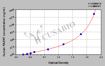
These standard curves are provided for demonstration only. A standard curve should be generated for each set of samples assayed.
ng/ml OD1 OD2 Average Corrected  
15 1.845 1.752 1.799 1.600  
7.5 1.532 1.476 1.504 1.305  
3.75 1.112 1.104 1.108 0.909  
1.88 0.784 0.739 0.762 0.563  
0.94 0.465 0.434 0.450 0.251  
0.47 0.376 0.359 0.368 0.169  
0.235 0.293 0.290 0.292 0.093  
0 0.201 0.197 0.199    
Troubleshooting
and FAQs
Storage
Store at 2-8°C. Please refer to protocol.
Lead Time
3-5 working days after you place the order, and it takes another 3-5 days for delivery via DHL or FedEx
Description

The Human Protein DJ-1 (PARK7) ELISA kit is the perfect solution for detection of the Parkinson disease protein 7 in human samples. This ELISA kit is ideal for researchers working in the field of cancer and nervous system disease research.

This ELISA kit is designed to detect PARK7 in serum, tissue homogenates, and cell lysates. With a detection range of 0.235 ng/mL-15 ng/mL and a sensitivity of 0.058 ng/mL, this kit provides precise and accurate results in just 1-5 hours.

Using the sandwich assay principle, this ELISA kit provides a quantitative measurement of PARK7 in your samples. With a sample volume of 50-100ul and a detection wavelength of 450 nm, this kit is both versatile and user-friendly.

The Human PARK7 ELISA kit has been validated with citations from over 3 publications, demonstrating its effectiveness in various research settings.

Customer Reviews and Q&A

 Customer Reviews

There are currently no reviews for this product.

Submit a Review here

Target Background

Function
(From Uniprot)
Multifunctional protein with controversial molecular function which plays an important role in cell protection against oxidative stress and cell death acting as oxidative stress sensor and redox-sensitive chaperone and protease. It is involved in neuroprotective mechanisms like the stabilization of NFE2L2 and PINK1 proteins, male fertility as a positive regulator of androgen signaling pathway as well as cell growth and transformation through, for instance, the modulation of NF-kappa-B signaling pathway. Has been described as a protein and nucleotide deglycase that catalyzes the deglycation of the Maillard adducts formed between amino groups of proteins or nucleotides and reactive carbonyl groups of glyoxals. But this function is rebuted by other works. As a protein deglycase, repairs methylglyoxal- and glyoxal-glycated proteins, and releases repaired proteins and lactate or glycolate, respectively. Deglycates cysteine, arginine and lysine residues in proteins, and thus reactivates these proteins by reversing glycation by glyoxals. Acts on early glycation intermediates (hemithioacetals and aminocarbinols), preventing the formation of advanced glycation endproducts (AGE) that cause irreversible damage. Also functions as a nucleotide deglycase able to repair glycated guanine in the free nucleotide pool (GTP, GDP, GMP, dGTP) and in DNA and RNA. Is thus involved in a major nucleotide repair system named guanine glycation repair (GG repair), dedicated to reversing methylglyoxal and glyoxal damage via nucleotide sanitization and direct nucleic acid repair. Protects histones from adduction by methylglyoxal, controls the levels of methylglyoxal-derived argininine modifications on chromatin. Able to remove the glycations and restore histone 3, histone glycation disrupts both local and global chromatin architecture by altering histone-DNA interactions as well as histone acetylation and ubiquitination levels. Displays a very low glyoxalase activity that may reflect its deglycase activity. Eliminates hydrogen peroxide and protects cells against hydrogen peroxide-induced cell death. Required for correct mitochondrial morphology and function as well as for autophagy of dysfunctional mitochondria. Plays a role in regulating expression or stability of the mitochondrial uncoupling proteins SLC25A14 and SLC25A27 in dopaminergic neurons of the substantia nigra pars compacta and attenuates the oxidative stress induced by calcium entry into the neurons via L-type channels during pacemaking. Regulates astrocyte inflammatory responses, may modulate lipid rafts-dependent endocytosis in astrocytes and neuronal cells. In pancreatic islets, involved in the maintenance of mitochondrial reactive oxygen species (ROS) levels and glucose homeostasis in an age- and diet dependent manner. Protects pancreatic beta cells from cell death induced by inflammatory and cytotoxic setting. Binds to a number of mRNAs containing multiple copies of GG or CC motifs and partially inhibits their translation but dissociates following oxidative stress. Metal-binding protein able to bind copper as well as toxic mercury ions, enhances the cell protection mechanism against induced metal toxicity. In macrophages, interacts with the NADPH oxidase subunit NCF1 to direct NADPH oxidase-dependent ROS production, and protects against sepsis.
Gene References into Functions
  1. DJ-1 protein concentration was significantly increased during the clinical phase in cerebrospinal fluid of sporadic Creutzfeldt-Jakob disease patients. PMID: 27975168
  2. The Parkinsonism-associated protein DJ-1 and its bacterial homologs deglycase 1/Hsp31, deglycase 2/YhbO, and deglycase 3/YajL function as deglycases that repair proteins and nucleotides from endogeneous glycation by glyoxal and methylglyoxal. DJ-1, deglycase 1 and deglycase 2 repair glyoxal- and methylglyoxal-glycated substrates, whereas deglycase 3 principally repairs glyoxal-glycated substrates. PMID: 29932913
  3. Data of this study showed that DJ-1 downregulation increased apoptosis and reduced migration by regulating matrix metalloproteinase 2 and matrix metalloproteinase 9 in HTR-8/SVneo cells under both ambient and oxidative stress. PMID: 29258409
  4. NRF2, DJ1 and SRNX1 are commonly expressed in diffusely infiltrating astrocytomas and they can be used in predicting patient prognosis. PMID: 29441509
  5. Low PARK7 expression is associated with pre-term birth. PMID: 29358306
  6. The results suggest that the impaired mitochondrial activity could be due to the broken interaction between DJ-1 and NDUFS3 and that downregulation of DJ-1 in sperm and testes contributes to AS pathogenesis. PMID: 29849492
  7. In this review, we discuss the role as well as the underlying molecular mechanisms of DJ-1 in ocular diseases, including Fuchs endothelial corneal dystrophy (FECD), age-related macular degeneration (AMD), cataracts, and ocular neurodegenerative diseases, highlighting that DJ-1 may serve as a very striking therapeutic target for ocular diseases. PMID: 29559831
  8. Novel candidate genes of the PARK7 interactome play a role in MS. PMID: 29100048
  9. results indicated that oxDJ-1 levels in erythrocytes can be used as a marker for the differential diagnosis of Parkinson's disease PMID: 28705587
  10. DJ-1 promoter polymorphism is associated with male infertility. PMID: 28932933
  11. The PD-associated L166P mutant, but not wild-type DJ-1, directly binds to and is cleaved by the mitochondrial serine protease Omi at both serine residues 3 and 121. PMID: 29177768
  12. DJ-1 plays a major role in various signaling pathways. Related to its anti-oxidant properties, it mediates cell survival and proliferation by activating the extracellular signal-regulated kinase (ERK1/2) pathway and attenuates cell death signaling by inhibiting apoptosis signal-regulating kinase 1 (ASK1) activation. [review] PMID: 28954246
  13. the present study highlighted the involvement of DJ1 in HGrelated EC injury and identified that DJ1 exerts a cellular protective effect in HUVECs exposed to HG induced oxidative stress via activation of the PI3K/AkteNOS signaling pathway. PMID: 29115508
  14. These findings suggest that oxidation-induced ubiquitination and degradation can be a quality control mechanism of oxidized redox-sensitive proteins including Prxs and DJ-1. PMID: 27703196
  15. study implies that DJ-1 may protect endothelial progenitor cells against Ang II-induced dysfunction by activating the PPARgamma/HO-1. PMID: 28600848
  16. Study shows the generation of oxDJ-1 in erythrocytes in early stage, unmedicated parkinson's disease (PD) patients and in non-human primates with PD symptoms. In addition, oxDJ-1 interacts with 20S proteasome in the erythrocytes of PD patients. The current findings suggest that oxidative stress is systemically present in PD patients and suggest that an early pathophysiological mechanism operates in PD. PMID: 27470541
  17. Not only is protein deglycase DJ-1 (DJ-1) linked to familial Parkinson's disease (PD), but it is also associated with the pathogenic mechanisms of sporadic PD and other neurodegenerative disorders where oxidative stress is implicated [Review]. PMID: 29147901
  18. Studies indicate that protein deglycase DJ-1 (DJ-1) plays a critical role in cancer [Review] PMID: 29147902
  19. Studies indicate that sperm protein deglycase DJ-1 (DJ-1 concentration in Asthenozoospermia is lower than that in control. PMID: 29147903
  20. Studies indicate Cys-106-oxidized protein deglycase DJ-1(oxDJ-1) levels in the brain and blood as a biomarker candidate for early-phase Parkinson's disease. PMID: 29147908
  21. Data suggest that protein deglycase DJ-1 (DJ-1) plays multiple roles in many cell types under metabolically challenging conditions as seen in obesity, insulin resistance, and type 2 diabetes mellitus [Review]. PMID: 29147909
  22. Studies suggest that protein deglycase DJ-1 (DJ-1) plays a critical role in tumor development and progression, and also as a therapeutic target against cancer. PMID: 29147911
  23. The clinical pattern of multisystem involvement and the biochemical findings in our patient highlight the role for DJ-1 in modulating mitochondrial response against oxidative stress. PMID: 27460976
  24. Oxidised DJ-1 was significantly decreased in idiopathic PD brain, suggesting altered complex function may also play a role in the more common sporadic form of the disease. PMID: 29016861
  25. High DJ1 expression is associated with breast cancer. PMID: 27582551
  26. Three types of pathogenic mutants of DJ-1 (M26I, D149A and L166P) have been reported to disrupt proper structures and lead to a loss of function--{REVIEW} PMID: 28458339
  27. the results suggest that over-oxidation may trigger loss of functions due to local structural modification in the Cys106 containing pocket of DJ-1 and structurally destabilize the dimeric state of DJ-1, which is believed to be its bioactive conformation. Such loss of functions would result in reduced ability of DJ-1 to protect from oxidative stress insults and may lead to increased progression of disease. PMID: 28844983
  28. This study demonstrated that the DJ1 mutations account for approximately 5% of EOPD patients from the Indian population. PMID: 27592010
  29. This report describe a new homozygous c.133C > T transition in DJ1 segregating with PD and ALS in a family from eastern Turkey. PMID: 26972524
  30. SiRNA-mediated DJ-1 knockdown reduced myeloma cell growth under a hypoxic microenvironment. PMID: 27734217
  31. chaperone-mediated autophagy protects mitochondrial function by degrading nonfunctional PARK7 and maintaining its homeostasis, and dysregulation of this pathway may contribute to the neuronal stress and death in Parkinson disease pathogenesis PMID: 27171370
  32. DJ-1 and its prokaryotic homologs constitute a major nucleotide repair system named guanine glycation repair. PMID: 28596309
  33. Taken together, our results provide a structural basis for the binding mechanism between DJ-1 and Bcl-XL, which will contribute to molecular understanding of the role of mitochondrial DJ-1 in Bcl-XL regulation in response to oxidative stress. PMID: 29175327
  34. Results suggest that overexpression of DJ-1 improves mitochondrial function, at least in part, through a mechanism involving Akt phosphorylation on threonine 308. PMID: 26913805
  35. Hsp31 is a homodimeric protein structurally similar to human DJ-1, a Parkinson's disease-linked protein, and both are members of the DJ-1/ThiJ/PfpI superfamily. PMID: 27097320
  36. DJ-1 co-aggregating with alpha-synuclein may result from weak hydrophobic interaction and DJ-1 exhibited chaperon-like activity in the initial time of alpha-synuclein aggregation at high temperature. PMID: 28330770
  37. The effects of variants in the Parkin, PINK1, and DJ-1 genes along with evidence for their pathogenicity have been summarized. (Review) PMID: 26965687
  38. genetic analyses of indel loci in ACE, DJ-1, and GIGYF2 genes, was performed to explore the potential contribution of insertion (I)/deletion (D) polymorphisms (indels) to the risk of PD in a Chinese population. PMID: 28873462
  39. DJ-1 deficiency negatively regulates liver progenitor cells (LPC) proliferation by impairing the formation of LPC-associated fibrosis and inflammatory niches. PMID: 27277679
  40. Downregulation of DJ-1 using DJ-1-siRNA in H69AR cells sensitized cancer cells to chemotherapeutic drugs through increasing drug-induced cell apoptosis accompanied with G0-G1 phase arrest. These findings suggest DJ-1 may serve as a potential biomarker for chemoresistance and prognostic factor for patients with small-cell lung cancer (SCLC). PMID: 28580701
  41. regulation of reactive oxygen species (ROS) and of the antioxidant protein DJ-1 (PARK-7), which increases with cancer progression and acts to lessen oxidative damage to malignant cells, in relationship with systemic mastocytosis severity, was examined. PMID: 27611333
  42. DJ-1 was highly expressed in the early stage of lung cancer, and its expression was significantly decreased after metastasis. Therefore, DJ-1 may be a potential biomarker for the early diagnosis and monitoring of lung cancer metastasis PMID: 28653888
  43. DJ-1 overexpression can restore the impaired Nrf2 pathway, leading to Alveolar type II cell protection in heavy smokers. PMID: 27093578
  44. DJ-1 serum levels seem to be correlated with worsening of the endometrial cancer grade and histotype, making it a reliable tool in the preoperative decision-making process. PMID: 28618925
  45. DJ-1 family deglycases repair proteins from glycation by glyoxals. Contradictory results suggested that this activity resulted from a Tris artefact. We show here that DJ-1 behaves indeed as a bona fide deglycase. PMID: 28013050
  46. ELISA-based detection of flotillin 2 and PARK7 showed that the combination of these proteins was able to distinguish prostate cancer patients and healthy controls with 68% sensitivity and 93% specificity PMID: 27664330
  47. Furthermore, SIRT1 activity was reduced in DJ-1-knockout cells, and this reduced activity was restored by re-introduction of wild-type DJ-1 but not of C106-mutant DJ-1 into DJ-1-knockout cells. It is first report showing direct connection of DJ-1 with SIRT1. PMID: 27105916
  48. DJ-1 is necessary for 20C-mediated protection against rotenone-induced oxidative damage, at least in part, by activating PI3K/Akt signaling, and subsequently enhancing the nuclear accumulation of Nrf2. PMID: 28245986
  49. The aberrant up-regulation of DJ-1 expression might be an important step in the pathogenesis of cervical carcinoma. PMID: 27544688
  50. Association of early onset Parkinson' and severe clinical signs with multiple alterations in DJ1 in Turkish patients. PMID: 27455133

Show More

Hide All

Involvement in disease
Parkinson disease 7 (PARK7)
Subcellular Location
Cell membrane; Lipid-anchor. Cytoplasm. Nucleus. Membrane raft. Mitochondrion. Endoplasmic reticulum.
Protein Families
Peptidase C56 family
Tissue Specificity
Highly expressed in pancreas, kidney, skeletal muscle, liver, testis and heart. Detected at slightly lower levels in placenta and brain (at protein level). Detected in astrocytes, Sertoli cells, spermatogonia, spermatids and spermatozoa. Expressed by panc
Database Links

HGNC: 16369

OMIM: 168600

KEGG: hsa:11315

STRING: 9606.ENSP00000340278

UniGene: Hs.419640

icon of phone
Call us
301-363-4651 (Available 9 a.m. to 5 p.m. CST from Monday to Friday)
icon of address
Address
7505 Fannin St., Ste 610, Room 7 (CUBIO Innovation Center), Houston, TX 77054, USA
icon of social media
Join us with

Subscribe newsletter

Leave a message

* To protect against spam, please pass the CAPTCHA test below.
CAPTCHA verification
© 2007-2024 CUSABIO TECHNOLOGY LLC All rights reserved. 鄂ICP备15011166号-1
webinars: DT3C facilitates antibody internalization X