Code | CSB-MP013481MOW |
Abbreviation | Recombinant Rhesus macaque MAPT protein (Active) |
MSDS | |
Size | $138 |
Order now | |
Image |
|
Have Questions? | Leave a Message or Start an on-line Chat |
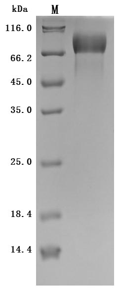
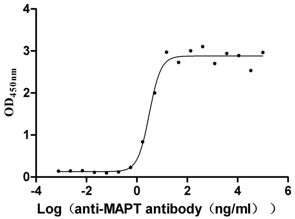
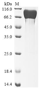
This recombinant microtubule-associated protein tau (MAPT) from Macaca mulatta is produced in mammalian cells with an N-terminal 10×His tag. Its expression region corresponds to the amino acids 2-459 of the Macaca mulatta MAPT protein. The recombinant MAPT protein exhibits high purity (>95% by SDS-PAGE) and meets strict endotoxin standards (<1.0 EU/μg, LAL method), ensuring research-grade quality for neuroscience applications.
Functional characterization confirms its biological activity through specific antibody binding in ELISA (EC50: 2.464-3.979 ng/mL against anti-MAPT antibody at 2 μg/mL immobilization). The mammalian expression system preserves critical post-translational modifications essential for MAPT protein function, including potential phosphorylation sites. The N-terminal 10×His tag facilitates purification while maintaining structural integrity. This recombinant MAPT protein serves as a valuable tool for investigating MAPT's role in microtubule stabilization, neurodegenerative mechanisms, and therapeutic development for tau-related disorders.
Macaca mulatta (rhesus macaque) serves as an important model organism for studying neurodegenerative diseases, particularly Alzheimer's disease (AD), and the role of microtubule-associated protein tau. Tau protein is integral to neuronal function, primarily by stabilizing microtubules within neuronal axons, a role that is vital for maintaining neuronal structure and function. In healthy neurons, tau exists in multiple isoforms generated through alternative splicing, which allows it to bind and stabilize microtubules effectively [1][2].
In the context of neurodegeneration, tau protein becomes hyperphosphorylated and forms pathological aggregates, leading to the development of neurofibrillary tangles, which are key hallmarks of Alzheimer's disease and other tauopathies [3-5]. Recent studies suggest that pathological tau conformers can be propagated from human to non-human primate brains, including in Macaca mulatta. This supports the notion that these animals can recapitulate facets of human tau pathology and may serve as a viable model for studying tau-related neurodegenerative processes [6].
Moreover, evidence indicates that tau pathology in Macaca mulatta can be influenced by amyloid-beta deposition. For instance, the intrathecal administration of amyloid-beta oligomers has been shown to elevate tau phosphorylation levels, thus mimicking aspects of Alzheimer's pathology [7]. Similarly, investigations have highlighted that the dura mater of Macaca mulatta displays significant anatomical and pathological features relevant to tauopathies, reinforcing the notion that these primates exhibit neurodegenerative disease characteristics akin to those seen in humans [8]. The ability to study tau dynamics and neurodegeneration in non-human primates adds depth to our understanding of human neurodegenerative diseases, potentially informing future therapeutic strategies [9].
References:
[1] J. Guo, S. Narasimhan, et al. Unique pathological tau conformers from alzheimer’s brains transmit tau pathology in nontransgenic mice. The Journal of Experimental Medicine, vol. 213, no. 12, p. 2635-2654, 2016. https://doi.org/10.1084/jem.20160833
[2] H. Kadavath, R. Hofele, et al. Tau stabilizes microtubules by binding at the interface between tubulin heterodimers. Proceedings of the National Academy of Sciences, vol. 112, no. 24, p. 7501-7506, 2015. https://doi.org/10.1073/pnas.1504081112
[3] J. Chang, H. Liu, et al. Effect of times to blood processing on the stability of blood proteins associated with dementia. Dementia and Geriatric Cognitive Disorders, vol. 49, no. 3, p. 303-311, 2020. https://doi.org/10.1159/000509358
[4] D. Beckman, P. Chakrabarty, et al. A novel tau‐based rhesus monkey model of alzheimer's pathogenesis. Alzheimer S & Dementia, vol. 17, no. 6, p. 933-945, 2021. https://doi.org/10.1002/alz.12318
[5] S. Narasimhan, J. Guo, et al. Pathological tau strains from human brains recapitulate the diversity of tauopathies in nontransgenic mouse brain. Journal of Neuroscience, vol. 37, no. 47, p. 11406-11423, 2017. https://doi.org/10.1523/jneurosci.1230-17.2017
[6] M. Darricau, C. Dou, et al. Tau seeds from alzheimer's disease brains trigger tau spread in macaques while oligomeric‐aβ mediates pathology maturation. Alzheimer S & Dementia, vol. 20, no. 3, p. 1894-1912, 2023. https://doi.org/10.1002/alz.13604
[7] D. Wakeman, M. Weed, et al. Intrathecal amyloid‐beta oligomer administration increases tau phosphorylation in the medial temporal lobe in the african green monkey: a nonhuman primate model of alzheimer's disease. Neuropathology and Applied Neurobiology, vol. 48, no. 4, 2022. https://doi.org/10.1111/nan.12800
[8] C. Janson, K. Hauser, et al. Characterization of non-human primate dura in health and neurodegeneration. 2022. https://doi.org/10.1101/2022.06.03.477517
[9] G. Stonebarger, H. Bimonte‐Nelson, & H. Urbanski. The rhesus macaque as a translational model for neurodegeneration and alzheimer’s disease. Frontiers in Aging Neuroscience, vol. 13, 2021. https://doi.org/10.3389/fnagi.2021.734173
Applications : Antigen
Review: I purchased lyophilized powder this time. After receiving the product, we dissolved it following the instructions, and it dissolved very well, and then through SDS detection, the purity was higher than the standard. After immunizing the mouse, coated the antigen, and several positive clones with good affinity were screened through ELISA. The antibody obtained from purified ascites has good specificity and high sensitivity. The overall performance of the product is good, the delivery is fast, and the COA report is very detailed!
By Anonymous