Recombinant Mouse Activin receptor type-2B (Acvr2b), partial (Active)

In Stock
Code CSB-MP001261MO1
Abbreviation Recombinant Mouse ACVR2B protein, partial (Active)
MSDS
Size $128
Order now
Image
  • (Tris-Glycine gel) Discontinuous SDS-PAGE (reduced) with 5% enrichment gel and 15% separation gel.
  • Activity
    Measured by its binding ability in a functional ELISA. Immobilized Mouse Acvr2b at 2 μg/ml can bind Anti-ACVR2A&ACVR2B recombinant antibody (CSB-RA623829MA1HU). The EC50 is 3.560-4.235 ng/mL. Biological Activity Assay
  • The purity of ACVR2B was greater than 90% as determined by SEC-HPLC
Have Questions? Leave a Message or Start an on-line Chat

Product Details

Purity
Greater than 95% as determined by SDS-PAGE.
Greater than 90% as determined by SEC-HPLC.
Endotoxin
Less than 1.0 EU/ug as determined by LAL method.
Activity
Measured by its binding ability in a functional ELISA. Immobilized Mouse Acvr2b at 2 μg/mL can bind Anti-ACVR2A&ACVR2B recombinant antibody (CSB-RA623829MA1HU). The EC50 is 3.560-4.235 ng/mL.
Target Names
Uniprot No.
Alternative Names
Activin receptor type-2B;EC: 2.7.11.30; Activin receptor type IIB (ACTR-IIB); Acvr2b
Species
Mus musculus (Mouse)
Source
Mammalian cell
Expression Region
19-137aa
Target Protein Sequence
SGRGEAETRECIYYNANWELERTNQSGLERCEGEQDKRLHCYASWRNSSGTIELVKKGCWLDDFNCYDRQECVATEENPQVYFCCCEGNFCNERFTHLPEPGGPEVTYEPPPTAPTLLT
Mol. Weight
15.2 kDa
Protein Length
Partial
Tag Info
C-terminal 10xHis-tagged
Form
Lyophilized powder
Note: We will preferentially ship the format that we have in stock, however, if you have any special requirement for the format, please remark your requirement when placing the order, we will prepare according to your demand.
Buffer
Lyophilized from a 0.2 μm filtered PBS, 6% Trehalose, pH 7.4
Reconstitution
We recommend that this vial be briefly centrifuged prior to opening to bring the contents to the bottom. Please reconstitute protein in deionized sterile water to a concentration of 0.1-1.0 mg/mL.We recommend to add 5-50% of glycerol (final concentration) and aliquot for long-term storage at -20℃/-80℃. Our default final concentration of glycerol is 50%. Customers could use it as reference.
Troubleshooting and FAQs
Storage Condition
Store at -20°C/-80°C upon receipt, aliquoting is necessary for mutiple use. Avoid repeated freeze-thaw cycles.
Shelf Life
The shelf life is related to many factors, storage state, buffer ingredients, storage temperature and the stability of the protein itself.
Generally, the shelf life of liquid form is 6 months at -20°C/-80°C. The shelf life of lyophilized form is 12 months at -20°C/-80°C.
Lead Time
3-7 business days
Notes
Repeated freezing and thawing is not recommended. Store working aliquots at 4℃ for up to one week.
Datasheet & COA
Please contact us to get it.
Description

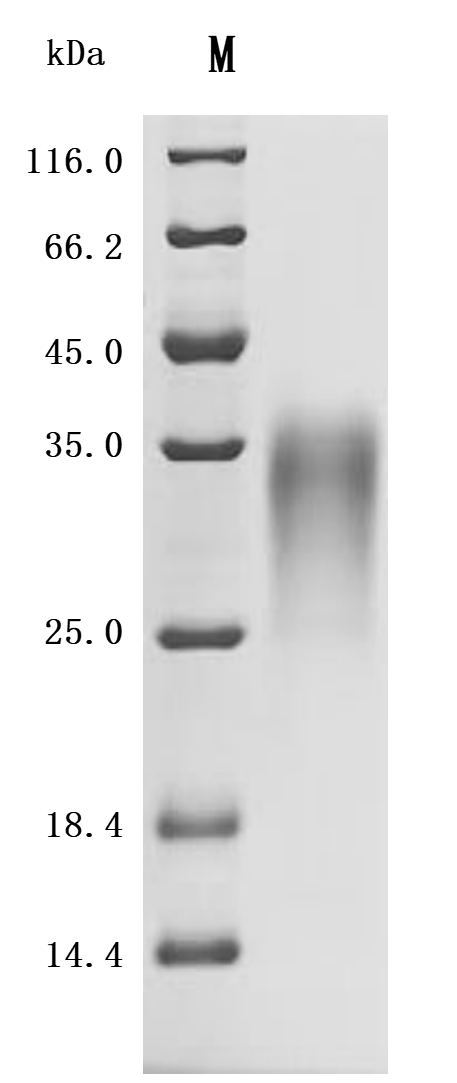
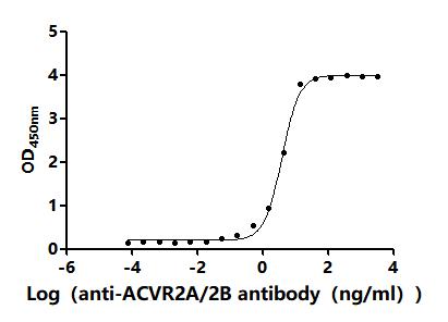
The recombinant mouse ACVR2B protein is co-expressed with a C-terminal 10*his-tag in mammalian cells. Its expression region encodes the 19-137aa of the mouse ACVR2B protein. It contains low endotoxin less than 1.0 EU/ug as measured by the LAL method. Its activity is validated through a functional ELISA where immobilized mouse ACVR2B at 2 μg/mL can bind the anti-ACVR2A&ACVR2B recombinant antibody (CSB-RA623829MA1HU), with the EC50 of 3.560-4.235 ng/mL.

The mouse ACVR2B protein is a member of the TGF-β superfamily of receptors. It plays a crucial role in various biological processes, particularly in muscle growth regulation and the response to muscle wasting conditions. ACVR2B functions primarily as a receptor for myostatin and other activins, which are negative regulators of muscle growth. The inhibition of ACVR2B signaling has been shown to lead to increased muscle mass, a phenomenon often referred to as double muscling in animals, including mice [1][2].

In the context of muscle physiology, ACVR2B is involved in the signaling pathways that mediate muscle atrophy and hypertrophy. Studies have demonstrated that transgenic mice expressing a dominant negative form of ACVR2B exhibit significant muscle hypertrophy due to the blockade of myostatin signaling [2][3]. The ACVR2B's activity is also critical in other systems, such as the heart, where its signaling is implicated in cardiac development and response to stress [4].

Moreover, ACVR2B has been implicated in pathological conditions such as cachexia, a syndrome characterized by muscle wasting often associated with cancer and other chronic diseases. Research indicates that blocking ACVR2B ligands can restore muscle protein synthesis and mitigate muscle loss in cachexia models [5][6]. The systemic administration of soluble forms of ACVR2B has been shown to improve muscle mass and function in various experimental settings, highlighting its potential as a therapeutic target for muscle-wasting disorders [7].

ACVR2B is also involved in developmental processes. Knockout studies have revealed that ACVR2B signaling is essential for proper vertebral development and overall embryonic growth, indicating its broader significance in developmental biology [8]. The receptor's interactions with various ligands, including activins and growth differentiation factors, further underscore its multifaceted role in cellular signaling and tissue homeostasis [9].

References:
[1] Y. Klimentidis, J. Bea, P. Thompson, W. Klimecki, C. Hu, G. Wu, et al. Genetic variant in acvr2b is associated with lean mass, Medicine & Science in Sports & Exercise, vol. 48, no. 7, p. 1270-1275, 2016. https://doi.org/10.1249/mss.0000000000000889
[2] S. Lee. Quadrupling muscle mass in mice by targeting tgf-ß signaling pathways, Plos One, vol. 2, no. 8, p. e789, 2007. https://doi.org/10.1371/journal.pone.0000789
[3] S. Lee, A. Lehar, J. Meir, C. Koch, A. Morgan, L. Warren, et al. Targeting myostatin/activin a protects against skeletal muscle and bone loss during spaceflight, Proceedings of the National Academy of Sciences, vol. 117, no. 38, p. 23942-23951, 2020. https://doi.org/10.1073/pnas.2014716117
[4] J. Hulmi, T. Nissinen, M. Räsänen, J. Degerman, J. Lautaoja, K. Hemanthakumar, et al. Prevention of chemotherapy‐induced cachexia by acvr2b ligand blocking has different effects on heart and skeletal muscle, Journal of Cachexia Sarcopenia and Muscle, vol. 9, no. 2, p. 417-432, 2017. https://doi.org/10.1002/jcsm.12265
[5] T. Nissinen, J. Hentilä, F. Penna, A. Lampinen, J. Lautaoja, V. Fachada, et al. Treating cachexia using soluble acvr2b improves survival, alters mtor localization, and attenuates liver and spleen responses, Journal of Cachexia Sarcopenia and Muscle, vol. 9, no. 3, p. 514-529, 2018. https://doi.org/10.1002/jcsm.12310
[6] T. Nissinen, J. Degerman, M. Räsänen, A. Poikonen, S. Koskinen, E. Mervaala, et al. Systemic blockade of acvr2b ligands prevents chemotherapy-induced muscle wasting by restoring muscle protein synthesis without affecting oxidative capacity or atrogenes, Scientific Reports, vol. 6, no. 1, 2016. https://doi.org/10.1038/srep32695
[7] D. DiGirolamo, V. Singhal, X. Chang, S. Lee, & E. Germain‐Lee. Administration of soluble activin receptor 2b increases bone and muscle mass in a mouse model of osteogenesis imperfecta, Bone Research, vol. 3, no. 1, 2015. https://doi.org/10.1038/boneres.2014.42
[8] Y. Lee, A. McPherron, S. Choe, Y. Sakai, R. Chandraratna, S. Lee, et al. Growth differentiation factor 11 signaling controls retinoic acid activity for axial vertebral development, Developmental Biology, vol. 347, no. 1, p. 195-203, 2010. https://doi.org/10.1016/j.ydbio.2010.08.022
[9] S. Lee, L. Reed, M. Davies, S. Girgenrath, M. Goad, K. Tomkinson, et al. Regulation of muscle growth by multiple ligands signaling through activin type ii receptors, Proceedings of the National Academy of Sciences, vol. 102, no. 50, p. 18117-18122, 2005. https://doi.org/10.1073/pnas.0505996102

Customer Reviews and Q&A

 Customer Reviews

There are currently no reviews for this product.

Submit a Review here

Target Background

Function
Transmembrane serine/threonine kinase activin type-2 receptor forming an activin receptor complex with activin type-1 serine/threonine kinase receptors (ACVR1, ACVR1B or ACVR1c). Transduces the activin signal from the cell surface to the cytoplasm and is thus regulating many physiological and pathological processes including neuronal differentiation and neuronal survival, hair follicle development and cycling, FSH production by the pituitary gland, wound healing, extracellular matrix production, immunosuppression and carcinogenesis. Activin is also thought to have a paracrine or autocrine role in follicular development in the ovary. Within the receptor complex, the type-2 receptors act as a primary activin receptors (binds activin-A/INHBA, activin-B/INHBB as well as inhibin-A/INHA-INHBA). The type-1 receptors like ACVR1B act as downstream transducers of activin signals. Activin binds to type-2 receptor at the plasma membrane and activates its serine-threonine kinase. The activated receptor type-2 then phosphorylates and activates the type-1 receptor. Once activated, the type-1 receptor binds and phosphorylates the SMAD proteins SMAD2 and SMAD3, on serine residues of the C-terminal tail. Soon after their association with the activin receptor and subsequent phosphorylation, SMAD2 and SMAD3 are released into the cytoplasm where they interact with the common partner SMAD4. This SMAD complex translocates into the nucleus where it mediates activin-induced transcription. Inhibitory SMAD7, which is recruited to ACVR1B through FKBP1A, can prevent the association of SMAD2 and SMAD3 with the activin receptor complex, thereby blocking the activin signal. Activin signal transduction is also antagonized by the binding to the receptor of inhibin-B via the IGSF1 inhibin coreceptor.
Gene References into Functions
  1. The current study reveals that ActRIIB activation by activin A induces muscle catabolism primarily through the activation of p38beta MAPK-mediated catabolic signalling that activates the ubiquitin-proteasome pathway and the autophagy-lysosome pathway. PMID: 27897407
  2. The endoplasmic reticulum stress stress and unfolded protein response are increased in mdx muscle. However, these processes are not distinctly improved by voluntary exercise or blocking activin receptor IIB ligands and thus do not appear to be optimal therapeutic choices for improving proteostasis in Duchenne muscular dystrophy. PMID: 27554968
  3. Data, including data from studies using transgenic mice, suggest that osteoblasts deficient in Acvr2A exhibit un-characteristic features; osteoclasts deficient in Acvr2A or Acvr2B or both appear normal. Acvr2A-deficient mice exhibit significantly increased femoral trabecular bone volume at 6 weeks of age; Acvr2B-deficient mice exhibit no significant change in any bone parameter. PMID: 28659341
  4. Differential muscle hypertrophy is associated with satellite cell numbers and Akt pathway activation following activin type IIB receptor inhibition in Mtm1 PMID: 24726641
  5. ActRIIB inhibition enhanced energy expenditure only at ambient temperature or in the cold, where nonshivering thermogenesis is minimal, suggesting that brown fat activation plays a prominent role in the metabolic actions of ActRIIB inhibition. PMID: 22586266
  6. findings best fit a model in which BMP3, produced by mature bone cells, acts to reduce BMP signaling through Acvr2b in skeletal progenitor cells, limiting their differentiation to mature osteoblasts PMID: 22074949
  7. Functional redundancy in osteoblast differentiation is observed between bone morphogenetic protein receptor BMPR-II and ActR-IIB. PMID: 21503889
  8. Inhibition of activin receptor type IIB increases strength and lifespan in myotubularin-deficient mice. PMID: 21281811
  9. Akt isoforms are not essential for for the ability of ActRIIB inhibition to regulate muscle size and function . PMID: 20856813
  10. ActRIIB plays a role in the specification of left-sidedness in developing mice PMID: 12112458
  11. Activin type IIB(ActRIIB) and its subfamily receptor, Activin type IIA (ActRIIA), cooperatively mediate the Gdf11 signal in patterning the axial vertebrae PMID: 12414726
  12. ActRIIB is expressed in the early development of thymocytes. PMID: 16477644
  13. genetic evidence strongly suggested that ActRIIB and Smad2 function in the same signaling pathway to regulate axial patterning and pancreas islet formation by means of a threshold mechanism. PMID: 17849440
  14. expression of MSTN and its associated binding proteins can be modulated in adipose tissue and skeletal muscle by chronic obesity PMID: 18334608
  15. Data suggest that BMP3 exerts its effects in the skeleton by altering signaling through ActRIIB in chondrocytes and the periosteum, and this results in defects in bone collar formation and late hypertrophic chondrocyte maturation. PMID: 19653325

Show More

Hide All

Subcellular Location
Cell membrane; Single-pass type I membrane protein.
Protein Families
Protein kinase superfamily, TKL Ser/Thr protein kinase family, TGFB receptor subfamily
Database Links
icon of phone
Call us
301-363-4651 (Available 9 a.m. to 5 p.m. CST from Monday to Friday)
icon of address
Address
7505 Fannin St., Ste 610, Room 7 (CUBIO Innovation Center), Houston, TX 77054, USA
icon of social media
Join us with

Subscribe newsletter

Leave a message

* To protect against spam, please pass the CAPTCHA test below.
CAPTCHA verification
© 2007-2025 CUSABIO TECHNOLOGY LLC All rights reserved. 鄂ICP备15011166号-1
Place an order now

I. Product details

*
*
*
*

II. Contact details

*
*

III. Ship To

*
*
*
*
*
*
*

IV. Bill To

*
*
*
*
*
*
*
*