Call us
301-363-4651 (Available 9 a.m. to 5 p.m. CST from Monday to Friday)
| Code | CSB-RA624111A0HU |
| Size | US$210 |
| Order now | |
| Image |
|
| Have Questions? | Leave a Message or Start an on-line Chat |
| Application | Recommended Dilution |
|---|---|
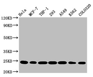
| WB | 1:500-1:5000 |
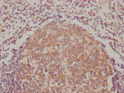
| IHC | 1:50-1:200 |
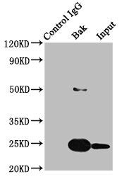
| IP | 1:200-1:1000 |
BAK1, also known as Bcl-2 homologous antagonist/killer, serves as a critical pro-apoptotic regulator within the intrinsic mitochondrial death pathway. As a member of the Bcl-2 protein family, BAK1 functions by permeabilizing the outer mitochondrial membrane upon activation, enabling cytochrome c release and subsequent caspase cascade initiation. Understanding BAK1 expression and regulation is essential for researchers investigating apoptotic mechanisms, cancer biology, and therapeutic strategies targeting programmed cell death.
This recombinant monoclonal antibody, generated against a synthetic peptide derived from human BAK1, offers the consistency and reliability that demanding experimental workflows require. As a sequence-defined recombinant clone, it eliminates the lot-to-lot variability inherent to traditional hybridoma-derived antibodies, ensuring reproducible results across long-term studies and multi-site collaborations.
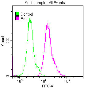
Validation across multiple applications demonstrates this antibody's experimental flexibility. Western blot analysis confirms specific detection of BAK1 at the predicted 24 kDa molecular weight across diverse human cell lines including MCF7, THP-1, HEK293, A431, and HepG2, with effective dilutions ranging from 1:500 to 1:5000. Immunohistochemical staining has been successfully performed in paraffin-embedded human endometrial tissue using citrate buffer antigen retrieval, while immunoprecipitation studies in HEK293 lysates confirm the antibody's capacity for protein enrichment applications. Flow cytometry validation in HeLa cells further extends its utility for single-cell analysis of intracellular BAK1 levels.
Whether investigating apoptotic signaling in cancer models, characterizing cell death pathways, or examining BAK1 expression patterns across tissues, this antibody provides researchers with a dependable tool for advancing cell biology research.
There are currently no reviews for this product.

