PCK2 Recombinant Monoclonal Antibody

Code CSB-RA900837A0HU
Size US$210
Order now
Image
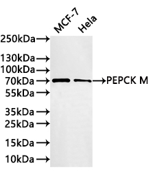
  • Western Blot
    Positive WB detected in: MCF-7 whole cell lysate(30µg), Hela whole cell lysate(30µg)
    All lanes: PEPCK M antibody at 1:1000
    Secondary
    Goat polyclonal to rabbit IgG at 1/40000 dilution
    Predicted band size: 71 kDa
    Observed band size: 71 kDa
    Exposure time:2min
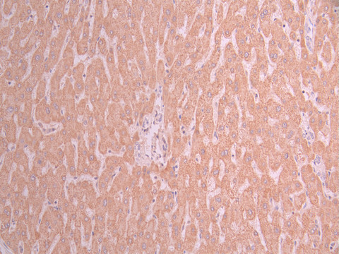
  • IHC image of CSB-RA900837A0HU diluted at 1:100 and staining in paraffin-embedded human liver cancer performed on a Leica BondTM system. After dewaxing and hydration, antigen retrieval was mediated by high pressure in a citrate buffer (pH 6.0). Section was blocked with 10% normal goat serum 30min at RT. Then primary antibody (1% BSA) was incubated at 4°C overnight. The primary is detected by a Goat anti-rabbit polymer IgG labeled by HRP and visualized using 0.05% DAB.
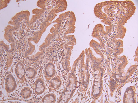
  • IHC image of CSB-RA900837A0HU diluted at 1:100 and staining in paraffin-embedded human small intestine tissue performed on a Leica BondTM system. After dewaxing and hydration, antigen retrieval was mediated by high pressure in a citrate buffer (pH 6.0). Section was blocked with 10% normal goat serum 30min at RT. Then primary antibody (1% BSA) was incubated at 4°C overnight. The primary is detected by a Goat anti-rabbit polymer IgG labeled by HRP and visualized using 0.05% DAB.
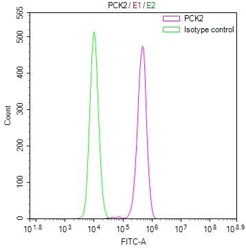
  • Overlay Peak curve showing Raji cells stained with CSB-RA900837A0HU (red line) at 1:100. The cells were fixed in 4% formaldehyde and permeated by 0.2% TritonX-100 for 10min. Then 10% normal goat serum to block non-specific protein-protein interactions followed by the antibody (1ug/1*106cells) for 45min at 4℃. The secondary antibody used was FITC-conjugated goat anti-rabbit IgG (H+L) at 1/200 dilution for 35min at 4℃.Control antibody (green line) was Rabbit IgG (1ug/1*106cells) used under the same conditions. Acquisition of >10,000 events was performed.
Have Questions? Leave a Message or Start an on-line Chat

Product Details

Uniprot No.
Target Names
Alternative Names
Phosphoenolpyruvate carboxykinase [GTP], mitochondrial (PEPCK-M) (EC 4.1.1.32), PCK2, PEPCK2
Species Reactivity
Human
Immunogen
A synthesized peptide from human PCK2 protein
Immunogen Species
Homo sapiens (Human)
Conjugate
Non-conjugated
Clonality
Monoclonal
Isotype
Rabbit IgG
Clone No.
3D12
Purification Method
Affinity-chromatography
Concentration
It differs from different batches. Please contact us to confirm it.
Buffer
Rabbit IgG in 10mM phosphate buffered saline , pH 7.4, 150mM sodium chloride, 0.05% BSA, 0.02% sodium azide and 50% glycerol.
Form
Liquid
Tested Applications
ELISA, WB, IHC, FC
Recommended Dilution
Application Recommended Dilution
WB 1:500-1:2000
IHC 1:50-1:200
FC 1:50-1:200
Troubleshooting and FAQs
Storage
Upon receipt, store at -20°C or -80°C. Avoid repeated freeze.
Lead Time
Basically, we can dispatch the products out in 1-3 working days after receiving your orders. Delivery time maybe differs from different purchasing way or location, please kindly consult your local distributors for specific delivery time.
Description

PCK2, also known as mitochondrial phosphoenolpyruvate carboxykinase (PEPCK-M), plays a critical role in gluconeogenesis and cellular energy metabolism. Unlike its cytosolic counterpart, this mitochondrial isoform catalyzes the GTP-dependent conversion of oxaloacetate to phosphoenolpyruvate within the mitochondrial matrix, positioning it as a key regulator of metabolic flux. Researchers investigating cancer metabolism, diabetes, and metabolic reprogramming increasingly recognize PCK2 as a significant target, given its emerging roles in tumor cell survival and metabolic adaptation.

This recombinant monoclonal antibody, generated from clone 3D12 in rabbit host, offers the reproducibility and consistency that demanding experimental workflows require. Because the antibody sequence is defined and production occurs through recombinant expression, you can expect reliable performance across experiments and between lots, eliminating the variability often encountered with traditional hybridoma-derived antibodies.

Validation data demonstrates robust performance across multiple platforms. Western blot analysis detects a clean 71 kDa band in both MCF-7 and HeLa whole cell lysates, matching the predicted molecular weight and confirming specificity for the target protein. Immunohistochemistry staining has been successfully performed in paraffin-embedded human liver cancer and small intestine tissues, revealing distinct localization patterns relevant to metabolic tissue studies. Flow cytometry validation using Raji cells shows clear positive signal separation from isotype controls, supporting intracellular detection applications.

Whether you are exploring metabolic enzyme expression in cancer cell lines, characterizing PCK2 distribution in human tissues, or quantifying protein levels across experimental conditions, this antibody provides the flexibility to move seamlessly between detection platforms while maintaining confidence in your results.

Usage
For Research Use Only. Not for use in diagnostic or therapeutic procedures.

Customer Reviews and Q&A

 Customer Reviews

There are currently no reviews for this product.

Submit a Review here

Target Background

Function
Catalyzes the conversion of oxaloacetate (OAA) to phosphoenolpyruvate (PEP), the rate-limiting step in the metabolic pathway that produces glucose from lactate and other precursors derived from the citric acid cycle.
Gene References into Functions
  1. Our study indicates that the T allele of the rs4982856 single-nucleotide polymorphisms in the PCK2 gene may be a risk factor for glucose intolerance after kidney transplantation PMID: 29731064
  2. Downregulation of PCK2 remodels tricarboxylic acid cycle in tumor-repopulating cells of melanoma PMID: 28166201
  3. ApoA-IV colocalizes with NR4A1, which suppresses G6Pase and PEPCK gene expression at the transcriptional level, reducing hepatic glucose output and lowering blood glucose. PMID: 26556724
  4. Results indicate that PEPCK promotes tumor growth by increasing glucose and glutamine metabolism, increases anabolic metabolism and promotes mTORC1 activity. PMID: 26481663
  5. Mitochondrial PCK2 regulates metabolic adaptation and enables glucose-independent tumor growth in various neoplasms. PMID: 26474064
  6. When autophagy was blocked, the level of glucose-6-phosphatase (G6Pase) and phosphoenolpyruvate carboxykinase (PEPCK) was reduced in HepG2 cells and not in Hep3B cells. PMID: 26036577
  7. PEPCK activity was elevated threefold in lung cancer samples over normal lungs and its activation mediates an adaptive response to glucose depletion in lung cancer. PMID: 24632615
  8. Amino acid limitation and ER stress inducers, conditions that activate the amino acid response (AAR) and the unfolded protein response (UPR), stimulate PCK2 gene transcription in tumor cell lines. PMID: 24973213
  9. Expression of phosphoenolpyruvate carboxykinase linked to chemoradiation susceptibility of human colon cancer cells. PMID: 24602180
  10. expression of HCV nonstructural component NS5A in Huh7 or primary hepatocytes stimulated PEPCK gene expression and glucose output in HepG2 cells. PMID: 22955269
  11. results reveal a novel link between glucose metabolism and the DNA damage signaling pathway and suggest a possible role for PEPCK and G6P in the DNA damage response PMID: 21733854
  12. Endoplasmic reticulum stress triggers suppression of AMPK while increasing C/EBPbeta and pCREB expression which activates PEPCK gene transcription. PMID: 20797423
  13. Results suggest that pepck2 gene expression is regulated by its 5' flanking region up to 822 bp, and 317 bp upstream of transcriptional start point. PMID: 15315819
  14. Wild-type AREBP, but not Ser(470) to Ala(470) substituted non-phosphorylating mutant, represses gene expression of the phosphoenolpyruvate carboxykinase (PEPCK), a key enzyme of gluconeogenesis. PMID: 17097062
  15. skeletal muscle PEPCK has a role in determining physical activity levels PMID: 19521512
  16. Increased transcriptional expression of PEPCK1 and G6Pc does not account for increased gluconeogenesis and fasting hyperglycemia in patients with type 2 diabetes mellitus. PMID: 19587243

Show More

Hide All

Involvement in disease
Mitochondrial phosphoenolpyruvate carboxykinase deficiency (M-PEPCKD)
Subcellular Location
Mitochondrion.
Protein Families
Phosphoenolpyruvate carboxykinase [GTP] family
Database Links

HGNC: 8725

OMIM: 261650

KEGG: hsa:5106

STRING: 9606.ENSP00000216780

UniGene: Hs.75812

icon of phone
Call us
301-363-4651 (Available 9 a.m. to 5 p.m. CST from Monday to Friday)

icon of address
Address
No. 269, Shendun 5th Road, Donghu Hi-Tech Development Area, Hubei Province, 430206, P.R.China
icon of social media
Join us with

Subscribe newsletter

Leave a message

* To protect against spam, please pass the CAPTCHA test below.
CAPTCHA verification
© 2007-2026 CUSABIO TECHNOLOGY LLC All rights reserved. 鄂ICP备15011166号-1
Place an order now

I. Product details

*
*
*
*

II. Contact details

*
*

III. Ship To

*
*
*
*
*
*
*

IV. Bill To

*
*
*
*
*
*
*
*