PLAA Recombinant Monoclonal Antibody

Code CSB-RA565057A0HU
Size US$210
Order now
Image
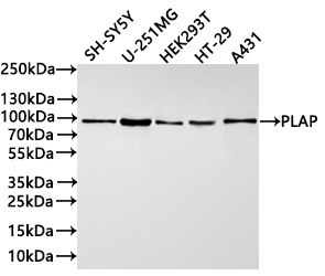
  • Western Blot
    Positive WB detected in: SH-SY5Y whole cell lysate(30µg), U-251MG whole cell lysate(30µg), HEK293T whole cell lysate(30µg), HT-29 whole cell lysate(30µg), A431 whole cell lysate(30µg)
    All lanes: PLAP antibody at 1:1000
    Secondary
    Goat polyclonal to rabbit IgG at 1/40000 dilution
    Predicted band size: 87 kDa
    Observed band size: 87 kDa
    Exposure time:1min40s
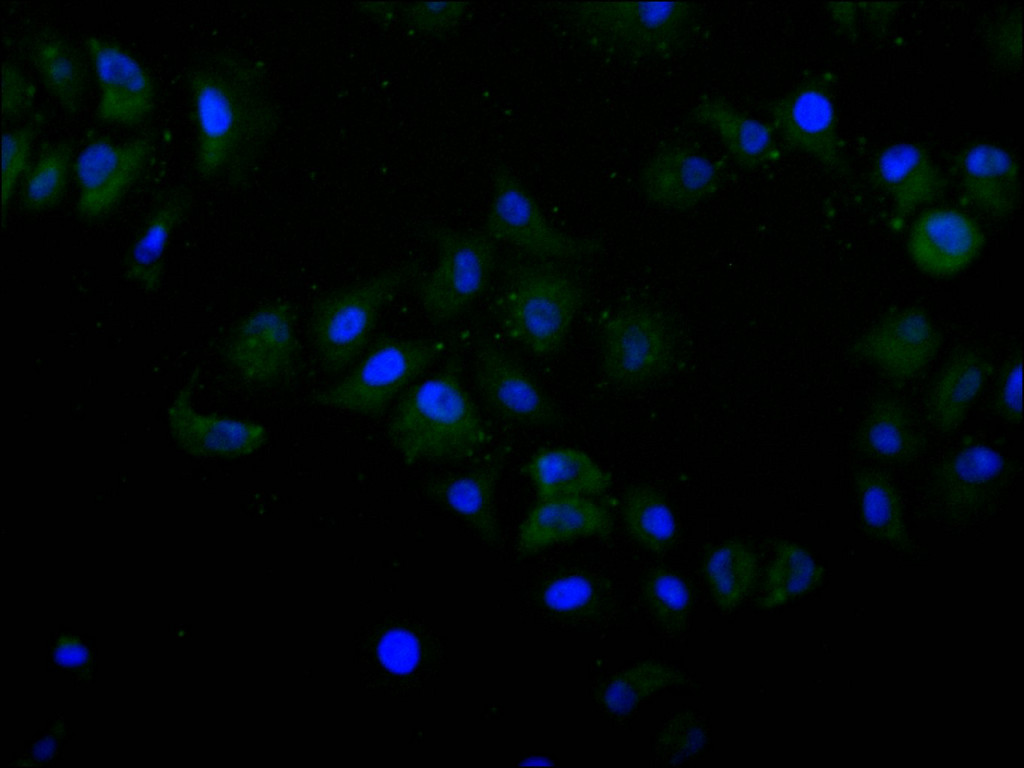
  • Immunofluorescence staining of Hela cell with CSB-RA565057A0HU at 1:50, counter-stained with DAPI. The cells were fixed in 4% formaldehyde, permeabilized using 0.2% Triton X-100 and blocked in 10% normal Goat Serum. The cells were then incubated with the antibody overnight at 4°C. The secondary antibody was Alexa Fluor 488-congugated AffiniPure Goat Anti-Rabbit IgG(H+L).
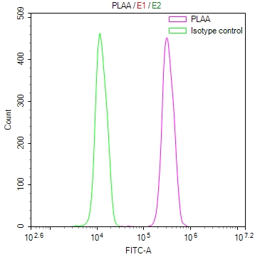
  • Overlay Peak curve showing Hela cells stained with CSB-RA565057A0HU (red line) at 1:100. The cells were fixed in 4% formaldehyde and permeated by 0.2% TritonX-100 for 10min. Then 10% normal goat serum to block non-specific protein-protein interactions followed by the antibody (1ug/1*106cells) for 45min at 4℃. The secondary antibody used was FITC-conjugated goat anti-rabbit IgG (H+L) at 1/200 dilution for 35min at 4℃.Control antibody (green line) was Rabbit IgG (1ug/1*106cells) used under the same conditions. Acquisition of >10,000 events was performed.
Have Questions? Leave a Message or Start an on-line Chat

Product Details

Uniprot No.
Target Names
Alternative Names
Phospholipase A-2-activating protein (PLA2P) (PLAP), PLAA, PLAP
Species Reactivity
Human
Immunogen
A synthesized peptide from human PLAA protein
Immunogen Species
Homo sapiens (Human)
Conjugate
Non-conjugated
Clonality
Monoclonal
Isotype
Rabbit IgG
Clone No.
1F3
Purification Method
Affinity-chromatography
Concentration
It differs from different batches. Please contact us to confirm it.
Buffer
Rabbit IgG in 10mM phosphate buffered saline , pH 7.4, 150mM sodium chloride, 0.05% BSA, 0.02% sodium azide and 50% glycerol.
Form
Liquid
Tested Applications
ELISA, WB, IF, FC
Recommended Dilution
Application Recommended Dilution
WB 1:500-1:2000
IF 1:50-1:200
FC 1:50-1:200
Troubleshooting and FAQs
Storage
Upon receipt, store at -20°C or -80°C. Avoid repeated freeze.
Lead Time
Basically, we can dispatch the products out in 1-3 working days after receiving your orders. Delivery time maybe differs from different purchasing way or location, please kindly consult your local distributors for specific delivery time.
Description

Phospholipase A-2-activating protein (PLAA) serves as a critical regulator of phospholipase A2 activity, positioning it at the intersection of lipid signaling, membrane dynamics, and cellular stress responses. This protein has garnered research attention for its roles in inflammatory pathways and its emerging connections to protein quality control mechanisms, making it a valuable target for investigators studying diverse cellular processes.

This recombinant monoclonal antibody, generated in rabbit against a synthetic peptide derived from human PLAA, offers the reproducibility and consistency that demanding experimental workflows require. Because the antibody sequence is defined and production occurs through recombinant technology, researchers can expect reliable performance across experiments and over time, eliminating the lot-to-lot variability that can complicate long-term studies.

Validation data demonstrates robust performance across multiple applications. Western blot analysis confirms specific detection of PLAA at the predicted 87 kDa molecular weight across a diverse panel of human cell lines, including neuroblastoma (SH-SY5Y), glioblastoma (U-251MG), embryonic kidney (HEK293T), colorectal adenocarcinoma (HT-29), and epidermoid carcinoma (A431) lysates. This broad detection profile across neural, epithelial, and other cell types provides flexibility for researchers working with various model systems. Immunofluorescence staining in HeLa cells reveals clear cytoplasmic localization patterns, while flow cytometry analysis demonstrates effective discrimination between specific signal and background, enabling quantitative single-cell studies.

The antibody's compatibility with ELISA, western blotting, immunofluorescence, and flow cytometry makes it well-suited for researchers investigating lipid signaling cascades, inflammatory responses, or protein homeostasis mechanisms where PLAA function may be implicated.

Usage
For Research Use Only. Not for use in diagnostic or therapeutic procedures.

Customer Reviews and Q&A

 Customer Reviews

There are currently no reviews for this product.

Submit a Review here

Target Background

Function
Plays a role in protein ubiquitination, sorting and degradation through its association with VCP. Involved in ubiquitin-mediated membrane proteins trafficking to late endosomes in an ESCRT-dependent manner, and hence plays a role in synaptic vesicle recycling. May play a role in macroautophagy, regulating for instance the clearance of damaged lysosomes. Plays a role in cerebellar Purkinje cell development. Positively regulates cytosolic and calcium-independent phospholipase A2 activities in a tumor necrosis factor alpha (TNF-alpha)- or lipopolysaccharide (LPS)-dependent manner, and hence prostaglandin E2 biosynthesis.
Gene References into Functions
  1. In both human and mouse, hypomorphic mutations in the ubiquitin adaptor protein PLAA cause an infantile-lethal neurodysfunction syndrome with seizures. PMID: 28413018
  2. This study presented that Phospholipase A2-activating protein is associated with a novel form of leukoencephalopathy. PMID: 28007986
  3. This review provides evidence that 1,25-dihydroxy vitamin D3 stimulates PLA2 via PLAA and CaMKII, a process initiated by Pdia3/PLAA interaction, which further triggers CaMKII-dependent PLA2 activation. [review] PMID: 25448737
  4. Function assignment to conserved residues in mammalian alkaline phosphatases. PMID: 11937510
  5. PLAP inhibits sPLA2-triggered release of fatty acids from erythrocyte membranes PMID: 14499668
  6. identified one stimulatory element, with Sp1 binding sites, and one inhibitory element, in exon 1 of the plaa gene PMID: 18291623
  7. Structural basis for ubiquitin recognition by a novel domain from the human phospholipase A2-activating protein PMID: 19423704

Show More

Hide All

Involvement in disease
Neurodevelopmental disorder with progressive microcephaly, spasticity, and brain anomalies (NDMSBA)
Subcellular Location
Nucleus. Cytoplasm. Cell junction, synapse.
Protein Families
WD repeat PLAP family
Database Links

HGNC: 9043

OMIM: 603873

KEGG: hsa:9373

STRING: 9606.ENSP00000380460

UniGene: Hs.27182

icon of phone
Call us
301-363-4651 (Available 9 a.m. to 5 p.m. CST from Monday to Friday)

icon of address
Address
No. 269, Shendun 5th Road, Donghu Hi-Tech Development Area, Hubei Province, 430206, P.R.China
icon of social media
Join us with

Subscribe newsletter

Leave a message

* To protect against spam, please pass the CAPTCHA test below.
CAPTCHA verification
© 2007-2026 CUSABIO TECHNOLOGY LLC All rights reserved. 鄂ICP备15011166号-1
Place an order now

I. Product details

*
*
*
*

II. Contact details

*
*

III. Ship To

*
*
*
*
*
*
*

IV. Bill To

*
*
*
*
*
*
*
*