Recombinant Human Interleukin-36 gamma protein (IL36G) (Active)

In Stock
Code CSB-AP002031HU
Abbreviation Recombinant Human IL36G protein (Active)
MSDS
Size $354
Order now
Image
Have Questions? Leave a Message or Start an on-line Chat
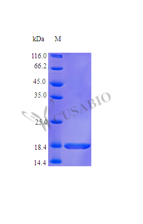
Product Details

Purity
>95% as determined by SDS-PAGE.
Endotoxin
Less than 1.0 EU/μg as determined by LAL method.
Activity
Fully biologically active when compared to standard. The specific activity is determined by its binding ability in a functional ELISA. Immobilized rHuIL-36γ at 1 ug/mL can bind recombinant human IL-1 Rrp2 Fc Chimera with a range of 0.15-5 ug/mL.
Target Names
IL36G
Uniprot No.
Research Area
Immunology
Alternative Names
IL 1 epsilon; IL 1 related protein 2; IL 1(EPSILON); IL 1F9; IL 1H1; IL 1RP2; IL-1 epsilon; IL-1-related protein 2; IL-1F9; IL-1H1; IL-1RP2; IL1E; Il1f9; IL1F9_HUMAN; IL1H1; IL1RP2; IL36 gamma; IL36G; Interleukin 1 epsilon; Interleukin 1 family member 9; Interleukin 1 homolog 1; Interleukin 1 related protein 2; Interleukin 36 gamma; Interleukin-1 epsilon; Interleukin-1 family member 9; Interleukin-1 homolog 1
Species
Homo sapiens (Human)
Source
E.coli
Expression Region
1-169aa
Complete Sequence
MRGTPGDADG GGRAVYQSMC KPITGTINDL NQQVWTLQGQ NLVAVPRSDS VTPVTVAVIT CKYPEALEQG RGDPIYLGIQ NPEMCLYCEK VGEQPTLQLK EQKIMDLYGQ PEPVKPFLFY RAKTGRTSTL ESVAFPDWFI ASSKRDQPII LTSELGKSYN TAFELNIND
Mol. Weight
18.7 kDa
Protein Length
Full Length
Tag Info
Tag-Free
Form
Lyophilized powder
Buffer
Lyophilized from a 0.2 µm filtered PBS, pH 7.4
Reconstitution
We recommend that this vial be briefly centrifuged prior to opening to bring the contents to the bottom. Please reconstitute protein in deionized sterile water to a concentration of 0.1-1.0 mg/mL.We recommend to add 5-50% of glycerol (final concentration) and aliquot for long-term storage at -20°C/-80°C. Our default final concentration of glycerol is 50%. Customers could use it as reference.
Troubleshooting and FAQs
Storage Condition
Store at -20°C/-80°C upon receipt, aliquoting is necessary for mutiple use. Avoid repeated freeze-thaw cycles.
Shelf Life
The shelf life is related to many factors, storage state, buffer ingredients, storage temperature and the stability of the protein itself.
Generally, the shelf life of liquid form is 6 months at -20°C/-80°C. The shelf life of lyophilized form is 12 months at -20°C/-80°C.
Lead Time
5-10 business days
Notes
Repeated freezing and thawing is not recommended. Store working aliquots at 4°C for up to one week.
Datasheet & COA
Please contact us to get it.
Description

Recombinant Human Interleukin-36 gamma protein (IL36G) is expressed in E. coli, covering the full-length sequence from 1 to 169 amino acids. This tag-free protein achieves high purity levels exceeding 95%, as confirmed by SDS-PAGE analysis. The protein appears to be biologically active, with specific activity validated through functional ELISA, demonstrating its ability to bind recombinant human IL-1 Rrp2 Fc Chimera. Endotoxin levels remain under 1.0 EU/µg, ensuring suitability for research applications.

Interleukin-36 gamma (IL-36γ) belongs to the interleukin-1 family and plays a role in immune response regulation. It participates in inflammatory pathways, contributing to the activation of immune cells such as T-cells and dendritic cells. Research on IL-36γ may be essential for understanding its function in immune processes and its potential implications in inflammatory diseases.

Potential Applications

Note: The applications listed below are based on what we know about this protein's biological functions, published research, and experience from experts in the field. However, we haven't fully tested all of these applications ourselves yet. We'd recommend running some preliminary tests first to make sure they work for your specific research goals.

1. IL-36γ/IL-1Rrp2 Receptor Binding Studies

This recombinant IL-36γ protein is confirmed to have receptor binding activity (binding to IL-1Rrp2 Fc chimera at 0.15-5 μg/mL) and is suitable for studying binding kinetics and affinity with its cognate receptor. Surface plasmon resonance, bio-layer interferometry, or ELISA-based assays can reliably characterize binding parameters. The high purity (>95%) and low endotoxin levels ensure minimal interference in sensitive binding assays. However, since activity validation is based solely on binding assays rather than functional cellular responses, researchers should confirm signaling competence in cellular assays for complete functional characterization.

2. Inflammatory Pathway Signaling Research

The protein can be used in cell-based assays studying inflammatory signaling, but its ability to activate downstream signaling pathways requires experimental confirmation. While the demonstrated receptor binding suggests potential signaling capability, researchers should first validate that this E. coli-expressed IL-36γ can indeed activate NF-κB, MAPK pathways, or cytokine production in relevant cell types before concluding inflammatory signaling mechanisms.

3. Antibody Development and Validation

This high-purity, tag-free IL-36γ protein serves as an excellent antigen for antibody development. The confirmed receptor binding activity indicates proper conformational epitopes, making it suitable for generating antibodies that recognize the native protein. However, antibodies should be validated for their ability to neutralize IL-36γ biological function using cellular assays, as binding activity alone doesn't guarantee functional neutralization capability.

4. Protein-Protein Interaction Screening

The protein can be used in interaction studies, but caution is needed when identifying novel binding partners beyond IL-1Rrp2. While the established IL-1Rrp2 binding provides a positive control, any newly identified interactors should be validated with additional methods, as E. coli expression lacks mammalian post-translational modifications that might affect some interactions.

5. Structural and Biophysical Characterization Studies

This protein is suitable for biophysical analyses (circular dichroism, dynamic light scattering) to assess folding and stability. However, for high-resolution structural studies (X-ray crystallography, cryo-EM), the protein may require optimization as E. coli expression might not yield the homogeneous conformation needed for crystallization. The receptor binding activity suggests proper folding, but structural studies would benefit from confirmation of monodispersity and stability under crystallization conditions.

Final Recommendation & Action Plan

This E. coli-expressed Human IL-36γ protein is a valuable tool for receptor binding studies and antibody development, with its biological activity confirmed through specific receptor interaction assays. For immediate use, prioritize binding kinetic studies with IL-1Rrp2 and antibody generation applications. For signaling studies, first validate the protein's ability to activate downstream pathways in relevant cell models before proceeding with extensive mechanistic research. For structural studies, perform initial biophysical characterization to assess suitability for high-resolution methods. While the demonstrated receptor binding indicates proper folding, researchers should note that the functional validation is limited to binding assays rather than cellular responses. For all applications, include appropriate controls and consider complementary experiments with mammalian-expressed IL-36γ when investigating aspects that might be affected by the absence of eukaryotic post-translational modifications.

Customer Reviews and Q&A

 Customer Reviews

There are currently no reviews for this product.

Submit a Review here

Target Background

Function
Cytokine that binds to and signals through the IL1RL2/IL-36R receptor which in turn activates NF-kappa-B and MAPK signaling pathways in target cells. Part of the IL-36 signaling system that is thought to be present in epithelial barriers and to take part in local inflammatory response; similar to the IL-1 system with which it shares the coreceptor IL1RAP. Seems to be involved in skin inflammatory response by acting on keratinocytes, dendritic cells and indirectly on T-cells to drive tissue infiltration, cell maturation and cell proliferation. In cultured keratinocytes induces the expression of macrophage, T-cell, and neutrophil chemokines, such as CCL3, CCL4, CCL5, CCL2, CCL17, CCL22, CL20, CCL5, CCL2, CCL17, CCL22, CXCL8, CCL20 and CXCL1; also stimulates its own expression and that of the prototypic cutaneous proinflammatory parameters TNF-alpha, S100A7/psoriasin and inducible NOS. May play a role in proinflammatory responses during particular neutrophilic airway inflammation: activates mitogen-activated protein kinases and NF-kappa B in primary lung fibroblasts, and stimulates the expression of IL-8 and CXCL3 and Th17 chemokine CCL20 in lung fibroblasts. May be involved in the innate immune response to fungal pathogens, such as Aspergillus fumigatus.
Gene References into Functions
  1. serum IL-36gamma levels were higher in active systemic lupus erythematosus patients and correlated with disease activity and arthritis PMID: 29571080
  2. Cathepsin S was identified as the major IL-36gamma-activating protease expressed in epithelial cells. PMID: 28289191
  3. enhanced expression of IL-36gamma was observed in plasma and bronchoalveolar lavage fluid of patients with acute respiratory distress syndrome because of bacterial pneumonia PMID: 28176791
  4. IL-36-mediated IL-6 and CXCL8 production in human lung fibroblasts and bronchial epithelial cells may be involved in pulmonary inflammation especially caused by bacterial or viral infections. PMID: 28869889
  5. With a focus on the skin as a target for microbial and viral invasion, the current knowledge of IL-36: IL-36alpha, IL-36beta and IL-36gamma, functions is reviewed. One physiological function of the IL-36smay be to counteract microbial immune evasion. [Review] PMID: 28811383
  6. IL-36gamma inhibits differentiation and induces inflammation of keratinocyte via Wnt signaling pathway in psoriasis. PMID: 28924372
  7. IL-36gamma-stimulated endothelial cells secrete the proinflammatory chemokines IL-8, CCL2 and CCL20. PMID: 27673278
  8. skin injury increases IL-36gamma via the activation of TLR3-SLUG-VDR axis and IL-36gamma induces REG3A to promote wound healing PMID: 28774595
  9. Autocrine and Paracrine Regulation of Keratinocyte Proliferation through a Novel Nrf2-IL-36gamma Pathway PMID: 27183581
  10. Here the authors show that Mycobacterium tuberculosis infection of macrophages induces IL-36gamma production in a 2-stage-regulated fashion. PMID: 27389350
  11. IL-36gamma, a member of the IL-1 superfamily, is involved in host defense and contributes to proinflammatory responses and development of inflammatory diseases. PMID: 27853811
  12. IL-36gamma is significantly more strongly expressed in the epidermis of patients with psoriasis-based erythroderma than in other inflammatory skin diseases. PMID: 26524325
  13. Study shows that plasma concentrations of IL-36alpha and IL-36gamma are overexpressed in active systemic lupus erythematosus patients and that IL-36alpha has a substantial pro-inflammatory effect through regulation of IL-6 and CXCL8 production. PMID: 26516833
  14. IRF6 is likely to promote inflammation to P. gingivalis through its regulation of IL-36gamma. PMID: 26819203
  15. Findings indicate that Interleukin (IL)-1beta-induced interleukin 36 gamma (IL-36gamma) expression is mediated by the activation of transcriptional factors, NF-kappaB p65 and AP-1 (c-jun). PMID: 26562662
  16. IL36G was identified as strong regulators of skin pathology in both lesional and non-lesional skin samples. PMID: 25897967
  17. Decreased Langerhans cell responses to IL36G: altered innate immunity in patients with recurrent respiratory papillomatosis PMID: 24950037
  18. IL-36gamma expression inversely correlated with the progression of human melanoma and lung cancer. PMID: 26321222
  19. IL-36gamma is a valuable biomarker in psoriasis patients, both for diagnostic purposes and measurement of disease activity during the clinical course PMID: 25525775
  20. CAMP induces IL-36gamma expression leading to initiation of skin inflammation and occasional exacerbations of psoriasis. PMID: 25305315
  21. IL-36 promotes myeloid cell infiltration, activation, and inflammatory activity in skin PMID: 24829417
  22. This is the first report of extracellular release of endogenous IL-36gamma through pyroptosis suggesting a function of IL-36gamma as an alarmin. PMID: 22318382
  23. Data presented herein shed further light on involvement of T-bet in innate immunity and suggest that IL-36gamma, besides IFNgamma, may contribute to functions of this transcription factor in immunopathology. PMID: 23095752
  24. Interleukin-36 (IL-36) ligands require processing for full agonist (IL-36alpha, IL-36beta, and IL-36gamma) or antagonist (IL-36Ra) activity PMID: 21965679
  25. Regulation and function of the IL-1 family cytokine IL-1F9 in human bronchial epithelial cells. PMID: 20870894
  26. Expression of IL-1F9 is increased in human plaque psoriasis skin and is overexpressed in a transgenic mouse psoriasis model. PMID: 21242515
  27. IL-1F6 and IL-1F8, in addition to IL-1F9, activate the pathway leading to NF-kappaB in an IL-1Rrp2-dependent manner in Jurkat cells PMID: 14734551
  28. This is the first report of IL-1 genotype association with the inflammation of skeletal muscle following acute resistance exercise that may potentially affect the adaptations to chronic resistance exercise. PMID: 15331687
  29. This report demonstrates expression of IL1F9 by bronchial epithelial cells induced by pro-inflammatory stimuli, suggesting a function of this molecule in airway inflammation. PMID: 15701729

Show More

Hide All

Subcellular Location
Cytoplasm. Secreted.
Protein Families
IL-1 family
Tissue Specificity
Highly expressed in tissues containing epithelial cells: skin, lung, stomach and esophagus. Expressed in bronchial epithelial. In skin is expressed only in keratinocytes but not in fibroblasts, endothelial cells or melanocytes. Up-regulated in lesional ps
Database Links

HGNC: 15741

OMIM: 605542

KEGG: hsa:56300

STRING: 9606.ENSP00000259205

UniGene: Hs.211238

icon of phone
Call us
301-363-4651 (Available 9 a.m. to 5 p.m. CST from Monday to Friday)
icon of address
Address
7505 Fannin St., Ste 610, Room 7 (CUBIO Innovation Center), Houston, TX 77054, USA
icon of social media
Join us with

Subscribe newsletter

Leave a message

* To protect against spam, please pass the CAPTCHA test below.
CAPTCHA verification
© 2007-2025 CUSABIO TECHNOLOGY LLC All rights reserved. 鄂ICP备15011166号-1
×
Place an order now

I. Product details

*
*
*
*

II. Contact details

*
*

III. Ship To

*
*
*
*
*
*
*

IV. Bill To

*
*
*
*
*
*
*
*