-
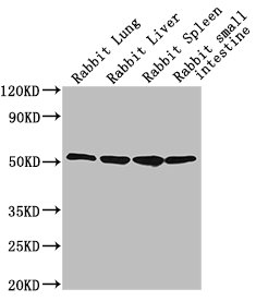
Western Blot
Positive WB detected in: Rabbit lung tissue, Rabbit liver tissue, Rabbit spleen tissue, Rabbit small intestine tissue
All lanes: CD14 antibody at 1:2500
Secondary
Goat polyclonal to Mouse IgG at 1/10000 dilution
Predicted band size: 41 kDa
Observed band size: 55 kDa
-
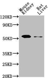
Western Blot
Positive WB detected in: Mouse kidney tissue, Rat liver tissue
All lanes: CD14 antibody at 1:2000
SecondaryGoat polyclonal to Mouse IgG at 1/10000 dilution
Predicted band size: 41 kDa
Observed band size: 55 kDa
-
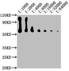
Western Blot
Positive WB detected in: NIH/3T3 whole cell lysate
All lanes: CD14 antibody at at 1:1000, 1:2000, 1:4000, 1:8000, 1:16000, 1:32000, 1:64000
Secondary
Goat polyclonal to Mouse IgG at 1/10000 dilution
Predicted band size: 41 kDa
Observed band size: 55 kDa
-
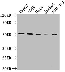
Western Blot
Positive WB detected in: HepG2 whole cell lysate, A549 whole cell lysate, Hela whole cell lysate, Jurkat whole cell lysate, NIH/3T3 whole cell lysate
All lanes: CD14 antibody at 1:1000
Secondary
Goat polyclonal to Mouse IgG at 1/10000 dilution
Predicted band size: 41 kDa
Observed band size: 55 kDa
-
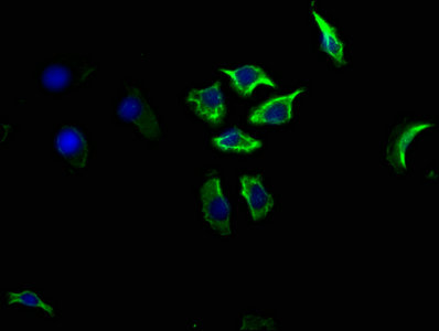
Immunofluorescence staining of A549 cells with CSB-MA004879A1m at 1:100, counter-stained with DAPI. The cells were blocked in 10% normal Goat Serum and then incubated with the primary antibody overnight at 4°C. The secondary antibody was Alexa Fluor 488-congugated AffiniPure Goat Anti-Mouse IgG(H+L).
-
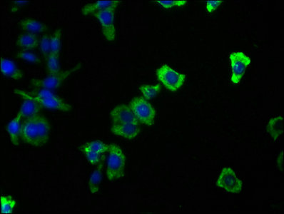
Immunofluorescence staining of HepG2 cells with CSB-MA004879A1m at 1:100, counter-stained with DAPI. The cells were blocked in 10% normal Goat Serum and then incubated with the primary antibody overnight at 4°C. The secondary antibody was Alexa Fluor 488-congugated AffiniPure Goat Anti-Mouse IgG(H+L).
-
Immunofluorescence staining of Raw264.7 cells with CSB-MA004879A1m at 1:100, counter-stained with DAPI. The cells were blocked in 10% normal Goat Serum and then incubated with the primary antibody overnight at 4°C. The secondary antibody was Alexa Fluor 488-congugated AffiniPure Goat Anti-Mouse IgG(H+L).