| Image |
-
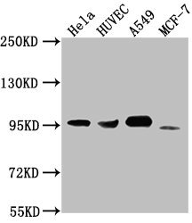
Western Blot
Positive WB detected in: Hela whole cell lysate, HUVEC whole cell lysate, A549 whole cell lysate, MCF-7 whole cell lysate
All lanes: CD44 antibody at 1:1500
Secondary
Goat polyclonal to Mouse IgG at 1/10000 dilution
Predicted band size: 82, 4, 78, 77, 81, 79, 75, 54,47, 40, 44, 33, 74, 76, 38, 16 kDa
Observed band size: 95 kDa
-
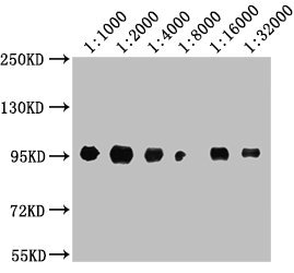
Western Blot
Positive WB detected in: A549 whole cell lysate
All lanes: CD44 antibody at 1:1000, 1:2000, 1:4000, 1:8000, 1:16000, 1:32000
Secondary
Goat polyclonal to Mouse IgG at 1/10000 dilution
Predicted band size: 82, 4, 78, 77, 81, 79, 75, 54,47, 40, 44, 33, 74, 76, 38, 16 kDa
Observed band size: 95 kDa
-
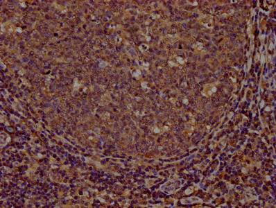
IHC image of CSB-MA004938A0m diluted at 1:100 and staining in paraffin-embedded human tonsil tissue performed on a Leica BondTM system. After dewaxing and hydration, antigen retrieval was mediated by high pressure in a citrate buffer (pH 6.0). Section was blocked with 10% normal goat serum 30min at RT. Then primary antibody (1% BSA) was incubated at 4°C overnight. The primary is detected by a biotinylated secondary antibody and visualized using an HRP conjugated SP system.
-
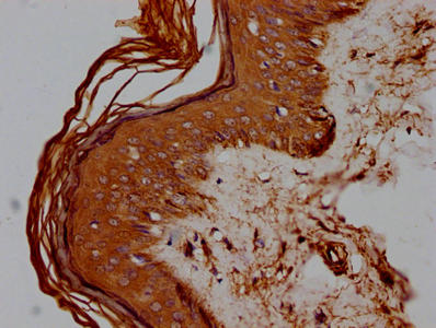
IHC image of CSB-MA004938A0m diluted at 1:100 and staining in paraffin-embedded human skin tissue performed on a Leica BondTM system. After dewaxing and hydration, antigen retrieval was mediated by high pressure in a citrate buffer (pH 6.0). Section was blocked with 10% normal goat serum 30min at RT. Then primary antibody (1% BSA) was incubated at 4°C overnight. The primary is detected by a biotinylated secondary antibody and visualized using an HRP conjugated SP system.
-
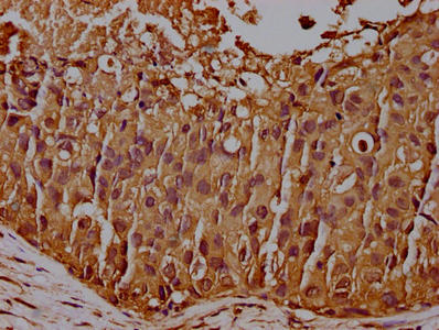
IHC image of CSB-MA004938A0m diluted at 1:100 and staining in paraffin-embedded human breast cancer performed on a Leica BondTM system. After dewaxing and hydration, antigen retrieval was mediated by high pressure in a citrate buffer (pH 6.0). Section was blocked with 10% normal goat serum 30min at RT. Then primary antibody (1% BSA) was incubated at 4°C overnight. The primary is detected by a biotinylated secondary antibody and visualized using an HRP conjugated SP system.
-
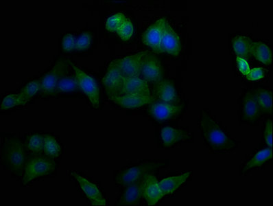
Immunofluorescence staining of Hela cells with CSB-MA004938A0m at 1:100, counter-stained with DAPI. The cells were blocked in 10% normal Goat Serum and then incubated with the primary antibody overnight at 4°C. The secondary antibody was Alexa Fluor 488-congugated AffiniPure Goat Anti-Mouse IgG(H+L).
-
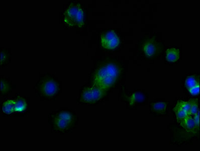
Immunofluorescence staining of MCF-7 cells with CSB-MA004938A0m at 1:100, counter-stained with DAPI. The cells were blocked in 10% normal Goat Serum and then incubated with the primary antibody overnight at 4°C. The secondary antibody was Alexa Fluor 488-congugated AffiniPure Goat Anti-Mouse IgG(H+L).
-
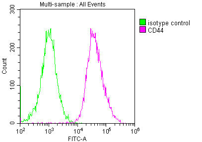
Overlay histogram showing Hela cells stained with CSB-MA004938A0m (red line) at 1:200. The cells were incubated in 1x PBS /10% normal goat serum to block non-specific protein-protein interactions followed by primary antibody for 1 h at 4°C. The secondary antibody used was FITC goat anti-mouse IgG(H+L) at 1/200 dilution for 1 h at 4°C. Isotype control antibody (green line) was used under the same conditions. Acquisition of >10,000 events was performed.
|