-
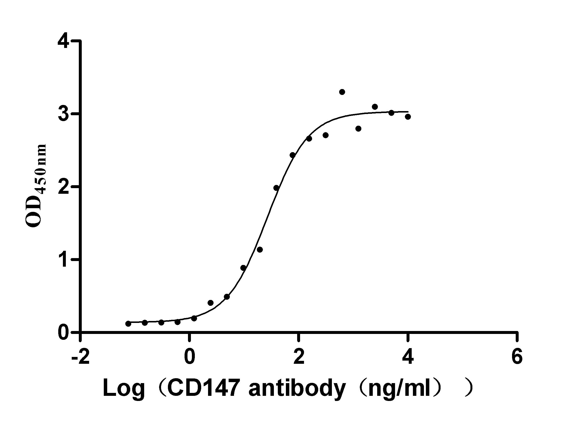
The Binding Activity of CD147 with Anti-CD147 recombinant Antibody
Activity: Measured by its binding ability in a functional ELISA. Immobilized Human CD147 (CSB-MP002831HU1) at 2 μg/ml can bind Anti-CD147 recombinant Antibody, the EC50 is 21.95-33.12 ng/ml.
-
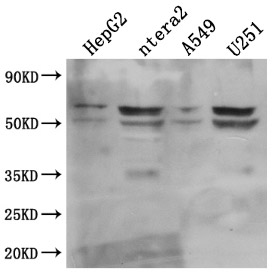
Western Blot
Positive WB detected in: HepG2 whole cell lysate, ntera2 whole cell lysate, A549 whole cell lysate, U251 whole cell lysate
All lanes: CD147 antibody at 1:1000
Secondary
Goat polyclonal to mouse IgG at 1/50000 dilution
Predicted band size: 42, 29, 23, 19 KDa
Observed band size: 35, 50-60 KDa
-
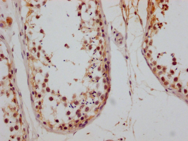
IHC image of CSB-RA002831A0HU diluted at 1:200 and staining in paraffin-embedded human testis tissue performed on a Leica BondTM system. After dewaxing and hydration, antigen retrieval was mediated by high pressure in a citrate buffer (pH 6.0). Section was blocked with 10% normal goat serum 30min at RT. Then primary antibody (1% BSA) was incubated at 4°C overnight. The primary is detected by a Goat anti-mouse polymer IgG labeled by HRP and visualized using 0.05% DAB.
-
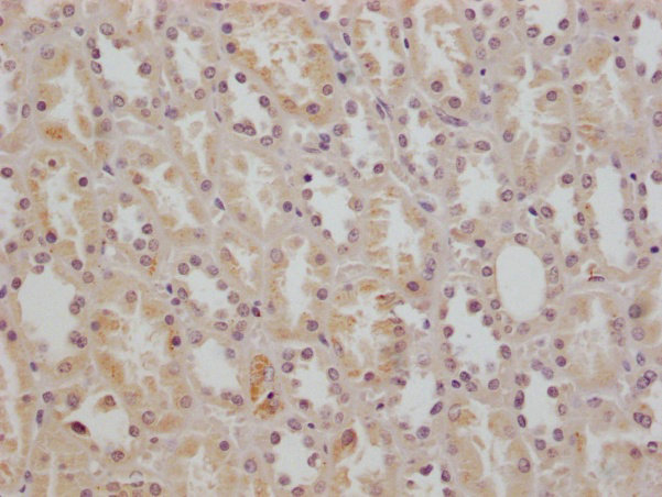
IHC image of CSB-RA002831A0HU diluted at 1:200 and staining in paraffin-embedded human kidney tissue performed on a Leica BondTM system. After dewaxing and hydration, antigen retrieval was mediated by high pressure in a citrate buffer (pH 6.0). Section was blocked with 10% normal goat serum 30min at RT. Then primary antibody (1% BSA) was incubated at 4°C overnight. The primary is detected by a Goat anti-mouse polymer IgG labeled by HRP and visualized using 0.05% DAB.
-
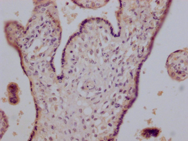
IHC image of CSB-RA002831A0HU diluted at 1:200 and staining in paraffin-embedded human placenta tissue performed on a Leica BondTM system. After dewaxing and hydration, antigen retrieval was mediated by high pressure in a citrate buffer (pH 6.0). Section was blocked with 10% normal goat serum 30min at RT. Then primary antibody (1% BSA) was incubated at 4°C overnight. The primary is detected by a Goat anti-mouse polymer IgG labeled by HRP and visualized using 0.05% DAB.
-
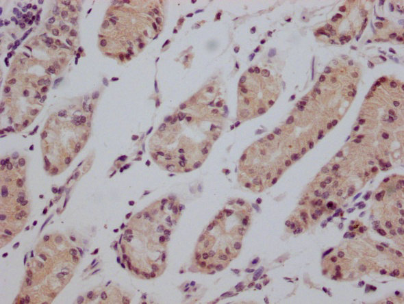
IHC image of CSB-RA002831A0HU diluted at 1:200 and staining in paraffin-embedded human stomach tissue performed on a Leica BondTM system. After dewaxing and hydration, antigen retrieval was mediated by high pressure in a citrate buffer (pH 6.0). Section was blocked with 10% normal goat serum 30min at RT. Then primary antibody (1% BSA) was incubated at 4°C overnight. The primary is detected by a Goat anti-mouse polymer IgG labeled by HRP and visualized using 0.05% DAB.
-
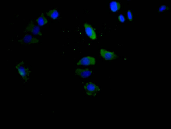
Immunofluorescence staining of Hela cells with CSB-RA002831A0HU at 1:150, counter-stained with DAPI. The cells were fixed in 4% formaldehyde and blocked in 10% normal Goat Serum. The cells were incubated with the antibody overnight at 4°C. Nuclear DNA was labeled in blue with DAPI. The secondary antibody was FITC-conjugated AffiniPure Goat Anti-Mouse IgG (H+L).
-
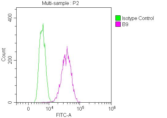
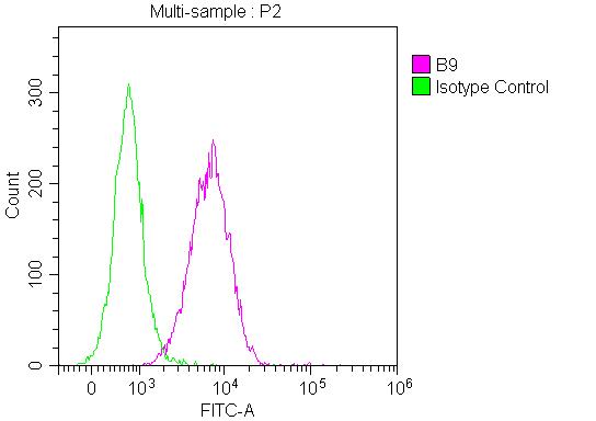
Overlay Peak curve showing Hela cells stained with CSB-RA002831A0HU (red line) with 1 μg/well (10 μg/mL, 100 μL/well). Then 10% normal goat serum was Incubated to block non-specific protein-protein interactions followed by the antibody (1µg/1*106cells) for 45 min at 4°C. The secondary antibody used was FITC-conjugated Goat Anti-Mouse IgG(H+L) at 1/200 dilution for 35 min at 4°C. Isotype control antibody (green line) was mouse IgG1 (1µg/1*106cells) used under the same conditions.Acquisition of >10,000 events was performed.
-
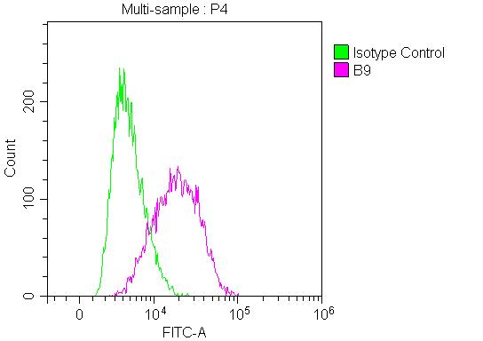
Overlay Peak curve showing Jurkat cells stained with CSB-RA002831A0HU (red line) with 1 μg/well (10 μg/mL, 100 μL/well). Then 10% normal goat serum was Incubated to block non-specific protein-protein interactions followed by the antibody (1µg/1*106cells) for 45 min at 4°C. The secondary antibody used was FITC-conjugated Goat Anti-Mouse IgG(H+L) at 1/200 dilution for 35 min at 4°C. Isotype control antibody (green line) was mouse IgG1 (1µg/1*106cells) used under the same conditions.Acquisition of >10,000 events was performed.
-
Overlay Peak curve showing HepG2 cells surface stained with CSB-RA002831A0HU (red line) with 1 μg/well (10 μg/mL, 100 μL/well). Then 10% normal goat serum was Incubated to block non-specific protein-protein interactions followed by the antibody (1µg/1*106cells) for 45 min at 4°C. The secondary antibody used was FITC-conjugated Goat Anti-Mouse IgG(H+L) at 1/200 dilution for 35 min at 4°C. Isotype control antibody (green line) was mouse IgG1 (1µg/1*106cells) used under the same conditions.Acquisition of >10,000 events was performed.