Call us
301-363-4651 (Available 9 a.m. to 5 p.m. CST from Monday to Friday)
| Code | CSB-RA009705A0HU |
| Size | US$210 |
| Order now | |
| Image |
|
| Have Questions? | Leave a Message or Start an on-line Chat |
| Application | Recommended Dilution |
|---|---|
| WB | 1:500-1:5000 |
| IF | 1:20-1:200 |
Glypican-3 is a heparan sulfate proteoglycan anchored to the cell surface that plays a critical role in regulating cell growth and differentiation during development. In adult tissues, GPC3 expression is typically silenced, but it becomes aberrantly reactivated in hepatocellular carcinoma, making it a valuable biomarker for liver cancer research and a promising therapeutic target. The protein also holds significance in stem cell biology, where it influences signaling pathways that govern cell fate decisions.
This recombinant monoclonal antibody, clone 4H11, offers the reproducibility and consistency that demanding research applications require. Because recombinant antibodies are produced from defined genetic sequences rather than hybridoma supernatants, you can expect uniform performance across experiments and over time, eliminating the lot-to-lot variability that can complicate long-term studies or multi-site collaborations.
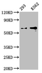
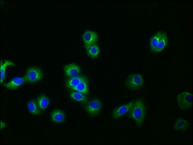
The antibody has been validated for Western blotting, immunofluorescence, and ELISA, giving you flexibility across different experimental workflows. In Western blot analysis, it successfully detects GPC3 in 293 and K562 whole cell lysates at working dilutions between 1:500 and 1:5000, producing a clear band at the expected 66 kDa molecular weight. For immunofluorescence applications, the antibody has been validated in HepG2 cells at dilutions from 1:20 to 1:200, demonstrating reliable performance in this hepatocellular carcinoma model that naturally expresses GPC3.
Supplied in a glycerol-containing buffer optimized for long-term stability, this antibody is well-suited for researchers investigating hepatocellular carcinoma biology, developmental signaling pathways, or stem cell regulation where consistent, sequence-defined detection of human GPC3 is essential.

Applications : Western Blot (WB)
Sample type: Jurkat whole cell lysate、Hep2B whole cell lysate/Human
Sample dilution: 1:1000
Review: I used CSB-RA009705A0HU antibody to conduct WB detection and detect the cell sample. The sample processing was as follows: After cracking with RIPA lysate, Protein loading Buffer was added to the sample, and the sample was cooked for 20min. The result was as follows: The product met the expectation.
By Anonymous

