Call us
301-363-4651 (Available 9 a.m. to 5 p.m. CST from Monday to Friday)
| Code | CSB-PA012474LA01HU |
| Size | US$166 |
| Order now | |
| Image |
|
| Promotion |  |
| Have Questions? | Leave a Message or Start an on-line Chat |
| Application | Recommended Dilution |
|---|---|
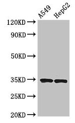
| WB | 1:500-1:2000 |
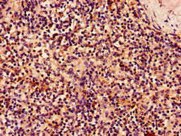
| IHC | 1:50-1:200 |
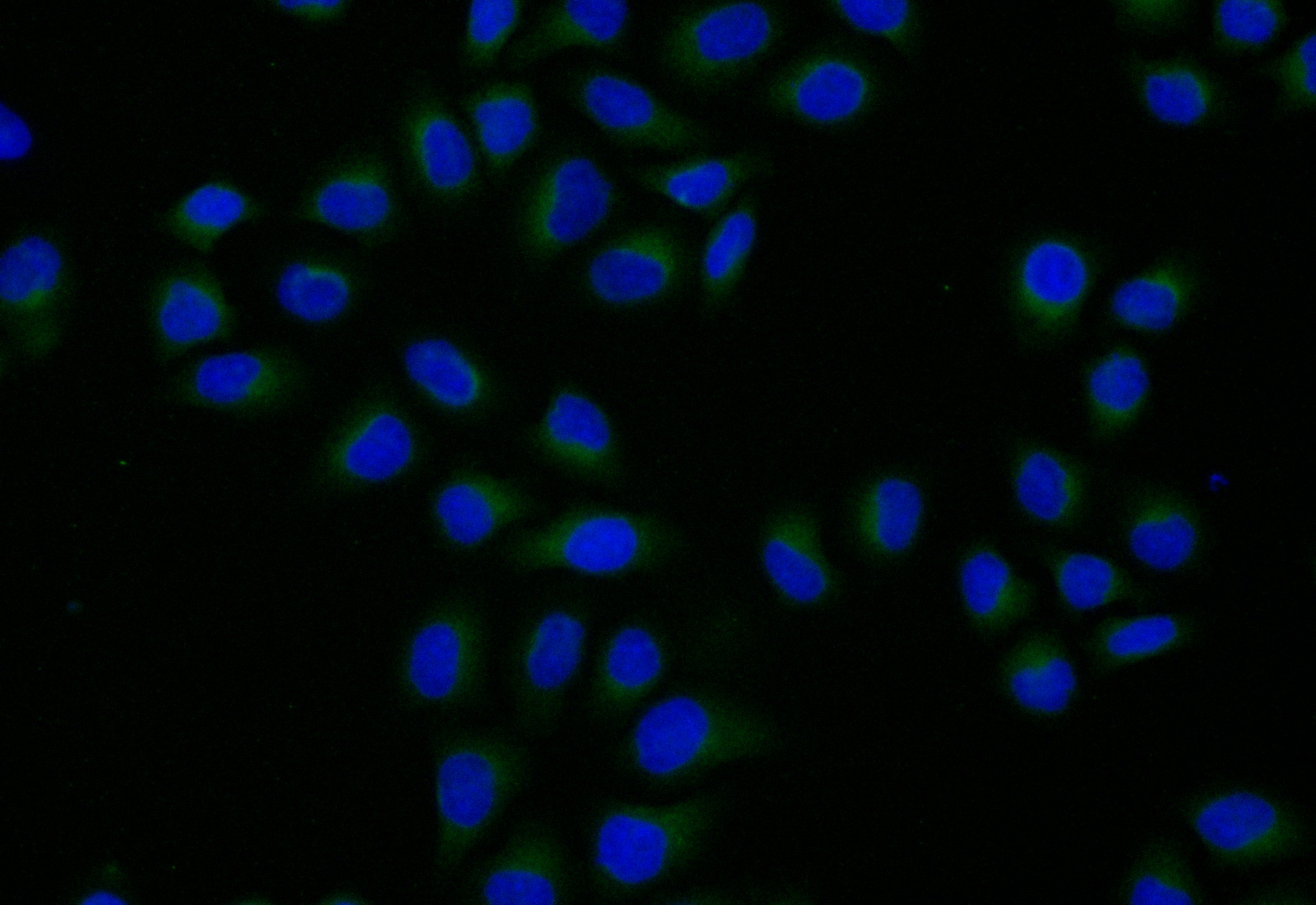
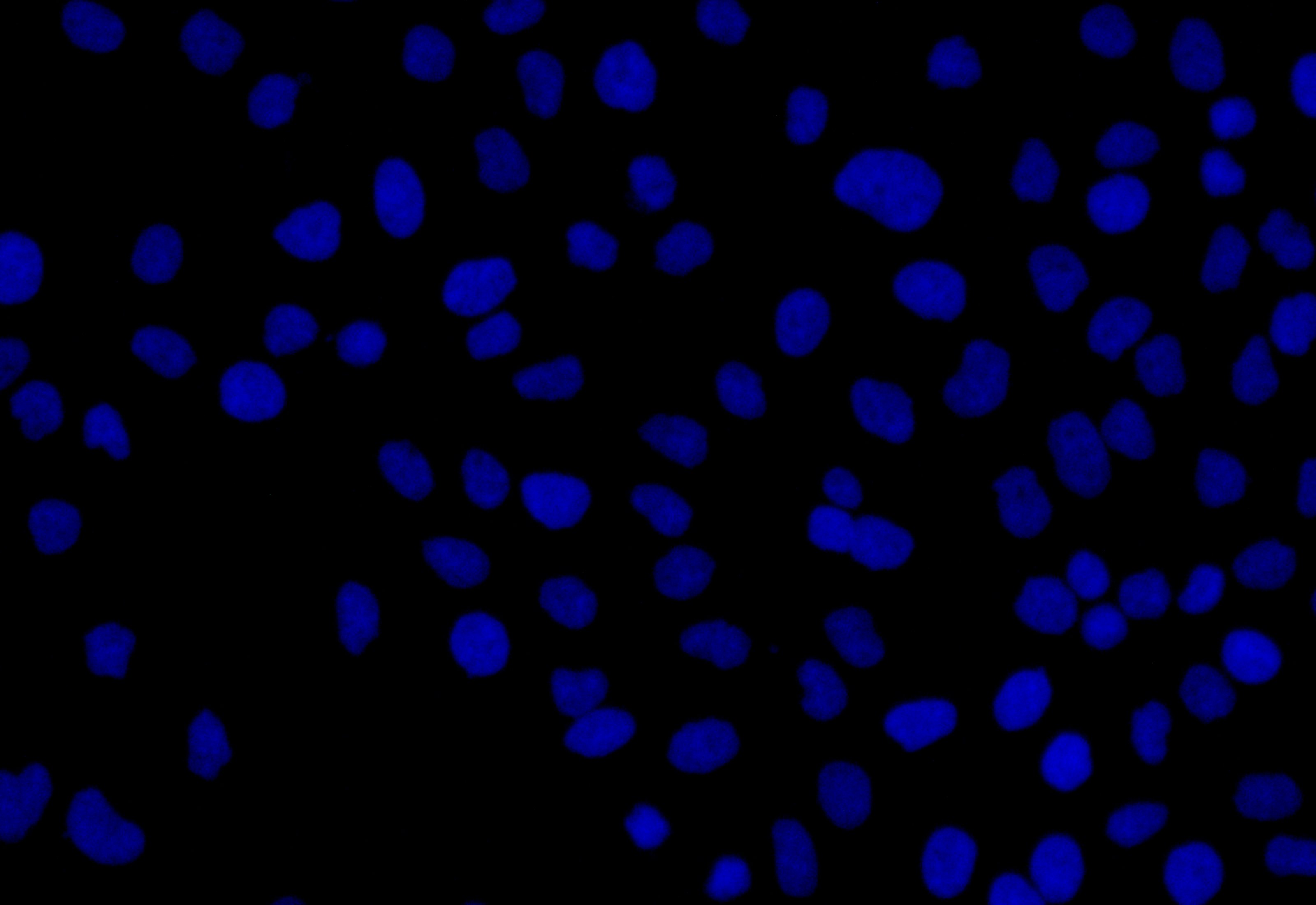
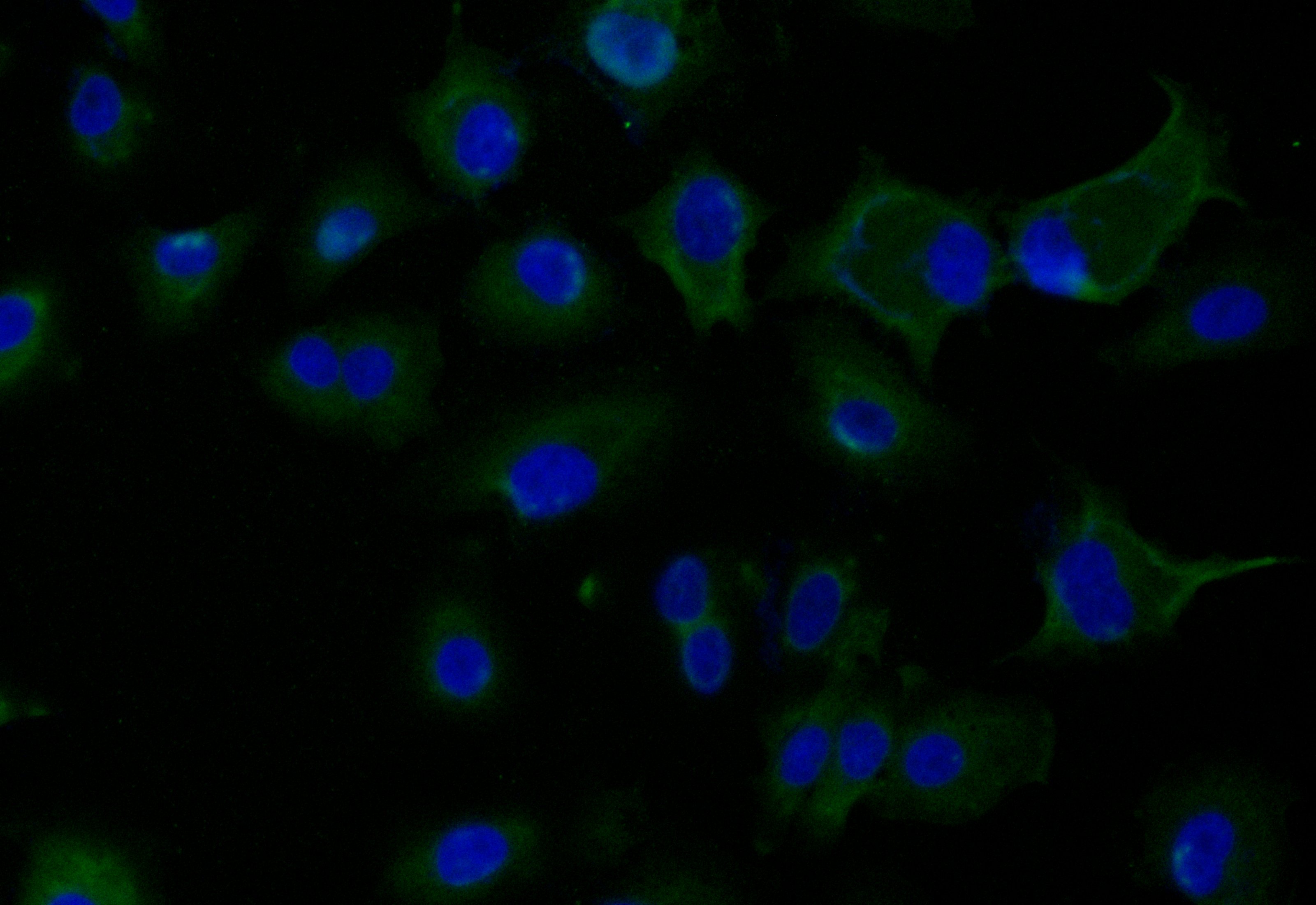
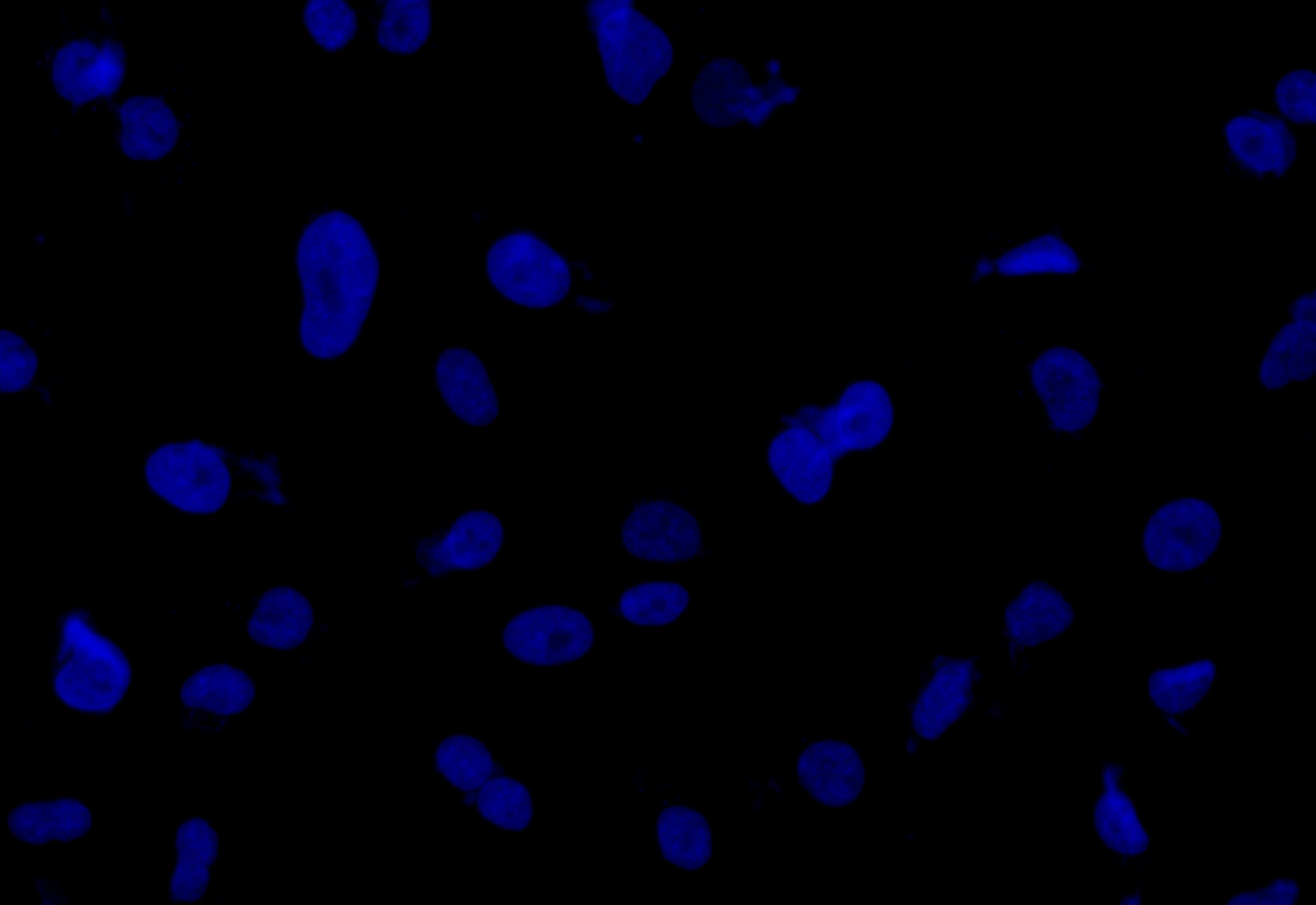
| IF | 1:20-1:100 |
The KLRK1 polyclonal antibody is a type of antibody that recognizes and binds to the human KLRK1 protein. The KLRK1 protein, also known as NKG2D, mainly functions to recognize and binds to stress-induced ligands expressed on the surface of infected or transformed cells, which allows the immune system to detect and eliminate these cells. KLRK1-mediated cytotoxicity is important for the immune response against viral infections and cancer, as well as for the elimination of cells undergoing cellular stress, such as cells undergoing DNA damage or apoptosis.
KLRK1 antibody is derived from multiple clones of B cells within the rabbit immunized with the recombinant human NKG2D protein (73-216aa), which produces a mixture of antibodies that can recognize different epitopes of the KLRK1 protein. The KLRK1 antibody underwent protein G affinity chromatography, getting a purity exceeding 95%. It has been quality validated in ELISA, WB, IHC, and IF applications.
There are currently no reviews for this product.

