Full Product Name
Rabbit anti-Homo sapiens (Human) FOS Polyclonal antibody
Alternative Names
Activator protein 1 antibody; AP 1 antibody; C FOS antibody; Cellular oncogene c fos antibody; Cellular oncogene fos antibody; FBJ murine osteosarcoma viral (v fos) oncogene homolog (oncogene FOS) antibody; FBJ murine osteosarcoma viral oncogene homolog antibody; FBJ murine osteosarcoma viral v fos oncogene homolog antibody; FBJ Osteosarcoma Virus antibody; FOS antibody; FOS protein antibody; FOS_HUMAN antibody; G0 G1 switch regulatory protein 7 antibody; G0/G1 switch regulatory protein 7 antibody; G0S7 antibody; Oncogene FOS antibody; p55 antibody; proto oncogene c Fos antibody; Proto oncogene protein c fos antibody; Proto-oncogene c-Fos antibody; v fos FBJ murine osteosarcoma viral oncogene homolog antibody
Species Reactivity
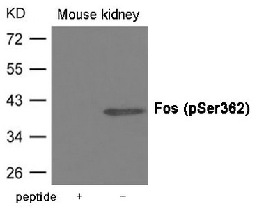
Human,Mouse,Rat
Immunogen
Peptide sequence around phosphorylation site of serine 362(K-G-S(p)-S-S) derived from Human Fos.
Immunogen Species
Homo sapiens (Human)
Purification Method
Antibodies were produced by immunizing rabbits with synthetic phosphopeptide and KLH conjugates. Antibodies were purified by affinity-chromatography using epitope-specific phosphopeptide. Non-phospho specific antibodies were removed by chromatogramphy usi
Concentration
It differs from different batches. Please contact us to confirm it.
Form
Supplied at 1.0mg/mL in phosphate buffered saline (without Mg2+ and Ca2+), pH 7.4, 150mM NaCl, 0.02% sodium azide and 50% glycerol.
Tested Applications
ELISA,WB
Recommended Dilution
| Application |
Recommended Dilution |
| WB |
1:500-1:1000 |
Storage
Upon receipt, store at -20°C or -80°C. Avoid repeated freeze.
Lead Time
Basically, we can dispatch the products out in 1-3 working days after receiving your orders. Delivery time maybe differs from different purchasing way or location, please kindly consult your local distributors for specific delivery time.
Usage
For Research Use Only. Not for use in diagnostic or therapeutic procedures.