Recombinant Mouse High mobility group protein B1 (Hmgb1), partial

In Stock
Code CSB-EP010553MO
Abbreviation Recombinant Mouse Hmgb1 protein, partial
MSDS
Size $388
Order now
Image
  • (Tris-Glycine gel) Discontinuous SDS-PAGE (reduced) with 5% enrichment gel and 15% separation gel.
Have Questions? Leave a Message or Start an on-line Chat

Product Details

Purity
Greater than 85% as determined by SDS-PAGE.
Target Names
Uniprot No.
Research Area
Epigenetics and Nuclear Signaling
Alternative Names
High mobility group protein 1
Species
Mus musculus (Mouse)
Source
E.coli
Expression Region
2-215aa
Target Protein Sequence
GKGDPKKPRGKMSSYAFFVQTCREEHKKKHPDASVNFSEFSKKCSERWKTMSAKEKGKFEDMAKADKARYEREMKTYIPPKGETKKKFKDPNAPKRPPSAFFLFCSEYRPKIKGEHPGLSIGDVAKKLGEMWNNTAADDKQPYEKKAAKLKEKYEKDIAAYRAKGKPDAAKKGVVKAEKSKKKKEEEDDEEDEEDEEEEEEEEDEDEEEDDDDE
Note: The complete sequence may include tag sequence, target protein sequence, linker sequence and extra sequence that is translated with the protein sequence for the purpose(s) of secretion, stability, solubility, etc.
If the exact amino acid sequence of this recombinant protein is critical to your application, please explicitly request the full and complete sequence of this protein before ordering.
Mol. Weight
25.7 kDa
Protein Length
Partial
Tag Info
N-terminal 6xHis-tagged
Form
Liquid or Lyophilized powder
Note: We will preferentially ship the format that we have in stock, however, if you have any special requirement for the format, please remark your requirement when placing the order, we will prepare according to your demand.
Buffer
If the delivery form is liquid, the default storage buffer is Tris/PBS-based buffer, 5%-50% glycerol.If the delivery form is lyophilized powder, the buffer before lyophilization is Tris/PBS-based buffer, 6% Trehalose.
Reconstitution
We recommend that this vial be briefly centrifuged prior to opening to bring the contents to the bottom. Please reconstitute protein in deionized sterile water to a concentration of 0.1-1.0 mg/mL.We recommend to add 5-50% of glycerol (final concentration) and aliquot for long-term storage at -20°C/-80°C. Our default final concentration of glycerol is 50%. Customers could use it as reference.
Troubleshooting and FAQs
Storage Condition
Store at -20°C/-80°C upon receipt, aliquoting is necessary for mutiple use. Avoid repeated freeze-thaw cycles.
Shelf Life
The shelf life is related to many factors, storage state, buffer ingredients, storage temperature and the stability of the protein itself.
Generally, the shelf life of liquid form is 6 months at -20°C/-80°C. The shelf life of lyophilized form is 12 months at -20°C/-80°C.
Lead Time
3-7 business days
Notes
Repeated freezing and thawing is not recommended. Store working aliquots at 4°C for up to one week.
Datasheet & COA
Please contact us to get it.
Description

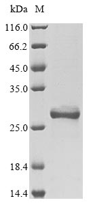
Recombinant Mouse High mobility group protein B1 (Hmgb1) is produced in E. coli and contains the amino acid sequence from 2 to 215. The protein is partially expressed with an N-terminal 6xHis-tag to make purification easier. SDS-PAGE analysis confirms it reaches a purity level of greater than 85%. This product is for research use only and should not be used in clinical applications.

High mobility group protein B1 (Hmgb1) is a non-histone chromosomal protein that appears to play a critical role in DNA architecture and regulation. The protein is involved in various cellular processes, including DNA repair, transcription, and replication. How Hmgb1 interacts with DNA and other nuclear proteins makes it an important subject when studying chromatin dynamics and gene expression.

Potential Applications

Note: The applications listed below are based on what we know about this protein's biological functions, published research, and experience from experts in the field. However, we haven't fully tested all of these applications ourselves yet. We'd recommend running some preliminary tests first to make sure they work for your specific research goals.

Based on the provided information, the recombinant mouse HMGB1 protein may not be correctly folded or fully bioactive without experimental validation. HMGB1 is a DNA-binding protein that requires proper folding of its two HMG boxes (A and B domains) for function. While E. coli expression systems can produce soluble HMGB1, the N-terminal 6xHis tag may sterically hinder the DNA-binding domains, as the DNA-binding sites are located near the N-terminus. The protein is partial length (2-215aa), which is nearly full-length but lacks the first amino acid; this might not significantly impact folding, but the tag could. The purity of >85% indicates the presence of impurities, including potential misfolded proteins or aggregates. Without validation (e.g., circular dichroism for secondary structure, DNA-binding assays like EMSA), the folding status and bioactivity remain uncertain.

1. Protein-DNA Interaction Studies

This recombinant mouse HMGB1 protein can be used to investigate DNA-binding properties only if correct folding and DNA-binding activity are experimentally verified. The N-terminal His-tag may interfere with DNA binding, leading to inaccurate results in SPR or EMSA assays. If misfolded, binding specificity studies may yield false negatives or altered kinetics. It is recommended to validate folding and DNA binding with positive controls (e.g., tag-free HMGB1) before use. The mouse origin is suitable for species-specific studies but does not guarantee functionality.

2. Antibody Development and Validation

This purified recombinant HMGB1 can serve as an antigen for antibody generation, but antibody specificity depends on the protein's folding state. If correctly folded, antibodies may recognize native epitopes; if misfolded, they could target non-conformational epitopes, reducing their utility for detecting physiological HMGB1. The His-tag may dominate the immune response, leading to tag-specific antibodies. For reliable outcomes, validate antibodies against full-length, tag-free HMGB1 or endogenous protein. The purity of>85% is acceptable but risks cross-reactivity with contaminants.

3. Protein-Protein Interaction Analysis

This HMGB1 protein can be used in pull-down assays only if folding is validated, as misfolding may cause non-specific interactions or false negatives. The partial length (2-215aa) retains major domains but may lack full interaction capabilities if the C-terminal region is critical. The His-tag allows immobilization but could promote tag-mediated artifacts. Include controls (e.g., tag-only protein) and verify folding with biophysical methods before interaction studies.

4. Structural and Biophysical Characterization

This recombinant HMGB1 is unsuitable for high-resolution structural studies (e.g., crystallography or NMR) without tag removal, as the His-tag introduces heterogeneity and flexibility. Biophysical techniques (e.g., CD, DLS) can assess general folding but may be confounded by the tag. The purity >85% is suboptimal for structural work (typically requiring >95%). For meaningful insights, remove the tag and confirm native-like structure through DNA-binding assays first.

5. In Vitro Functional Assays

This HMGB1 protein can be used in chromatin remodeling assays only if bioactivity is confirmed. The His-tag may affect DNA bending or nucleosome positioning, leading to misleading functional data. If misfolded, results may not reflect physiological HMGB1 behavior. Validate activity with known assays (e.g., nucleosome sliding) and compare with tag-free standards. The mouse sequence is appropriate for mouse-derived systems but requires folding validation.

Final Recommendation & Action Plan

To ensure reliable outcomes, first validate the folding and bioactivity of the recombinant HMGB1 using techniques such as circular dichroism to confirm expected secondary structure (e.g., alpha-helical content in HMG boxes), electrophoretic mobility shift assays (EMSA) to verify DNA-binding capability, and size-exclusion chromatography to assess oligomeric state and purity. If possible, remove the 6xHis tag via proteolytic cleavage and re-purify the tag-free protein for functional and structural studies. For applications like antibody development, proceed with caution and validate antibodies thoroughly. Always include appropriate controls, such as tag-free HMGB1 or DNA-binding mutants, to account for potential artifacts. Given the E. coli expression system, while it can produce functional HMGB1, independent validation is critical due to the tag and purity concerns.

Customer Reviews and Q&A

 Customer Reviews

There are currently no reviews for this product.

Submit a Review here

Target Background

Function
Multifunctional redox sensitive protein with various roles in different cellular compartments. In the nucleus is one of the major chromatin-associated non-histone proteins and acts as a DNA chaperone involved in replication, transcription, chromatin remodeling, V(D)J recombination, DNA repair and genome stability. Proposed to be an universal biosensor for nucleic acids. Promotes host inflammatory response to sterile and infectious signals and is involved in the coordination and integration of innate and adaptive immune responses. In the cytoplasm functions as sensor and/or chaperone for immunogenic nucleic acids implicating the activation of TLR9-mediated immune responses, and mediates autophagy. Acts as danger associated molecular pattern (DAMP) molecule that amplifies immune responses during tissue injury. Released to the extracellular environment can bind DNA, nucleosomes, IL-1 beta, CXCL12, AGER isoform 2/sRAGE, lipopolysaccharide (LPS) and lipoteichoic acid (LTA), and activates cells through engagement of multiple surface receptors. In the extracellular compartment fully reduced HMGB1 (released by necrosis) acts as a chemokine, disulfide HMGB1 (actively secreted) as a cytokine, and sulfonyl HMGB1 (released from apoptotic cells) promotes immunological tolerance. Has proangiogenic activity. May be involved in platelet activation. Binds to phosphatidylserine and phosphatidylethanolamide. Bound to RAGE mediates signaling for neuronal outgrowth. May play a role in accumulation of expanded polyglutamine (polyQ) proteins.; Nuclear functions are attributed to fully reduced HGMB1. Associates with chromatin and binds DNA with a preference to non-canonical DNA structures such as single-stranded DNA, DNA-containing cruciforms or bent structures, supercoiled DNA and ZDNA. Can bent DNA and enhance DNA flexibility by looping thus providing a mechanism to promote activities on various gene promoters by enhancing transcription factor binding and/or bringing distant regulatory sequences into close proximity. May be involved in nucleotide excision repair (NER), mismatch repair (MMR) and base excision repair (BER) pathways, and double strand break repair such as non-homologous end joining (NHEJ). Involved in V(D)J recombination by acting as a cofactor of the RAG complex: acts by stimulating cleavage and RAG protein binding at the 23 bp spacer of conserved recombination signal sequences (RSS). In vitro can displace histone H1 from highly bent DNA. Can restructure the canonical nucleosome leading to relaxation of structural constraints for transcription factor-binding. Enhances binding of sterol regulatory element-binding proteins (SREBPs) such as SREBF1 to their cognate DNA sequences and increases their transcriptional activities. Facilitates binding of TP53 to DNA. Proposed to be involved in mitochondrial quality control and autophagy in a transcription-dependent fashion implicating HSPB1; however, this function has been questioned. Can modulate the activity of the telomerase complex and may be involved in telomere maintenance.; In the cytoplasm proposed to dissociate the BECN1:BCL2 complex via competitive interaction with BECN1 leading to autophagy activation. Can protect BECN1 and ATG5 from calpain-mediated cleavage and thus proposed to control their proautophagic and proapoptotic functions and to regulate the extent and severity of inflammation-associated cellular injury. In myeloid cells has a protective role against endotoxemia and bacterial infection by promoting autophagy. Involved in endosomal translocation and activation of TLR9 in response to CpG-DNA in macrophages.; In the extracellular compartment (following either active secretion or passive release) involved in regulation of the inflammatory response. Fully reduced HGMB1 (which subsequently gets oxidized after release) in association with CXCL12 mediates the recruitment of inflammatory cells during the initial phase of tissue injury; the CXCL12:HMGB1 complex triggers CXCR4 homodimerization. Induces the migration of monocyte-derived immature dendritic cells and seems to regulate adhesive and migratory functions of neutrophils implicating AGER/RAGE and ITGAM. Can bind to various types of DNA and RNA including microbial unmethylated CpG-DNA to enhance the innate immune response to nucleic acids. Proposed to act in promiscuous DNA/RNA sensing which cooperates with subsequent discriminative sensing by specific pattern recognition receptors. Promotes extracellular DNA-induced AIM2 inflammasome activation implicating AGER/RAGE. Disulfide HMGB1 binds to transmembrane receptors, such as AGER/RAGE, TLR2, TLR4 and probably TREM1, thus activating their signal transduction pathways. Mediates the release of cytokines/chemokines such as TNF, IL-1, IL-6, IL-8, CCL2, CCL3, CCL4 and CXCL10. Promotes secretion of interferon-gamma by macrophage-stimulated natural killer (NK) cells in concert with other cytokines like IL-2 or IL-12. TLR4 is proposed to be the primary receptor promoting macrophage activation and signaling through TLR4 seems to implicate LY96/MD-2. In bacterial LPS- or LTA-mediated inflammatory responses binds to the endotoxins and transfers them to CD14 for signaling to the respective TLR4:LY96 and TLR2 complexes. Contributes to tumor proliferation by association with ACER/RAGE. Can bind to IL1-beta and signals through the IL1R1:IL1RAP receptor complex. Binding to class A CpG activates cytokine production in plasmacytoid dendritic cells implicating TLR9, MYD88 and AGER/RAGE and can activate autoreactive B cells. Via HMGB1-containing chromatin immune complexes may also promote B cell responses to endogenous TLR9 ligands through a B-cell receptor (BCR)-dependent and ACER/RAGE-independent mechanism. Inhibits phagocytosis of apoptotic cells by macrophages; the function is dependent on poly-ADP-ribosylation and involves binding to phosphatidylserine on the cell surface of apoptotic cells. In adaptive immunity may be involved in enhancing immunity through activation of effector T-cells and suppression of regulatory T (TReg) cells. In contrast, without implicating effector or regulatory T-cells, required for tumor infiltration and activation of T-cells expressing the lymphotoxin LTA:LTB heterotrimer thus promoting tumor malignant progression. Also reported to limit proliferation of T-cells. Released HMGB1:nucleosome complexes formed during apoptosis can signal through TLR2 to induce cytokine production. Involved in induction of immunological tolerance by apoptotic cells; its pro-inflammatory activities when released by apoptotic cells are neutralized by reactive oxygen species (ROS)-dependent oxidation specifically on Cys-106. During macrophage activation by activated lymphocyte-derived self apoptotic DNA (ALD-DNA) promotes recruitment of ALD-DNA to endosomes.
Subcellular Location
Nucleus. Cytoplasm. Secreted. Chromosome. Cell membrane; Peripheral membrane protein; Extracellular side. Endosome. Endoplasmic reticulum-Golgi intermediate compartment.
Protein Families
HMGB family
Tissue Specificity
Serum levels are found elevated in mice with modeled systemic lupus erythematosus (SLE) and are correlated with SLE disease activity.
Database Links
icon of phone
Call us
301-363-4651 (Available 9 a.m. to 5 p.m. CST from Monday to Friday)
icon of address
Address
7505 Fannin St., Ste 610, Room 7 (CUBIO Innovation Center), Houston, TX 77054, USA
icon of social media
Join us with

Subscribe newsletter

Leave a message

* To protect against spam, please pass the CAPTCHA test below.
CAPTCHA verification
© 2007-2025 CUSABIO TECHNOLOGY LLC All rights reserved. 鄂ICP备15011166号-1
×
Place an order now

I. Product details

*
*
*
*

II. Contact details

*
*

III. Ship To

*
*
*
*
*
*
*

IV. Bill To

*
*
*
*
*
*
*
*