PKN2 Recombinant Monoclonal Antibody

Code CSB-RA921617A0HU
Size US$210
Order now
Image
  • Western Blot
    Positive WB detected in: Hela whole cell lysate, U251 whole cell lysate, PC3 whole cell lysate, MCF-7 whole cell lysate
    All lanes: PKN2 antibody at 1:2000
    Secondary
    Goat polyclonal to rabbit IgG at 1/50000 dilution
    Predicted band size: 113, 117, 112 kDa
    Observed band size: 130 kDa
  • IHC image of CSB-RA921617A0HU diluted at 1:100 and staining in paraffin-embedded human colon cancer performed on a Leica BondTM system. After dewaxing and hydration, antigen retrieval was mediated by high pressure in a citrate buffer (pH 6.0). Section was blocked with 10% normal goat serum 30min at RT. Then primary antibody (1% BSA) was incubated at 4°C overnight. The primary is detected by a Goat anti-rabbit polymer IgG labeled by HRP and visualized using 0.05% DAB.
  • Immunofluorescence staining of MCF-7 cell with CSB-RA921617A0HU at 1:50, counter-stained with DAPI. The cells were fixed in 4% formaldehyde and blocked in 10% normal Goat Serum. The cells were then incubated with the antibody overnight at 4°C. The secondary antibody was Alexa Fluor 509-congugated AffiniPure Goat Anti-Rabbit IgG(H+L).
  • Overlay Peak curve showing MCF7 cells stained with CSB-RA921617A0HU (red line) at 1:100. The cells were fixed in 4% formaldehyde and permeated by 0.2% TritonX-100. Then 10% normal goat serum to block non-specific protein-protein interactions followed by the antibody (1ug/1*106cells) for 45min at 4℃. The secondary antibody used was FITC-conjugated Goat Anti-rabbit IgG(H+L) at 1:200 dilution for 35min at 4℃.Control antibody (green line) was rabbit IgG (1ug/1*106cells) used under the same conditions. Acquisition of >10,000 events was performed.
Have Questions? Leave a Message or Start an on-line Chat
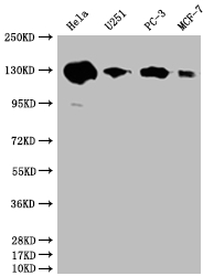
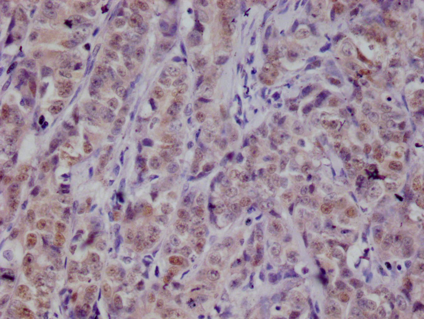
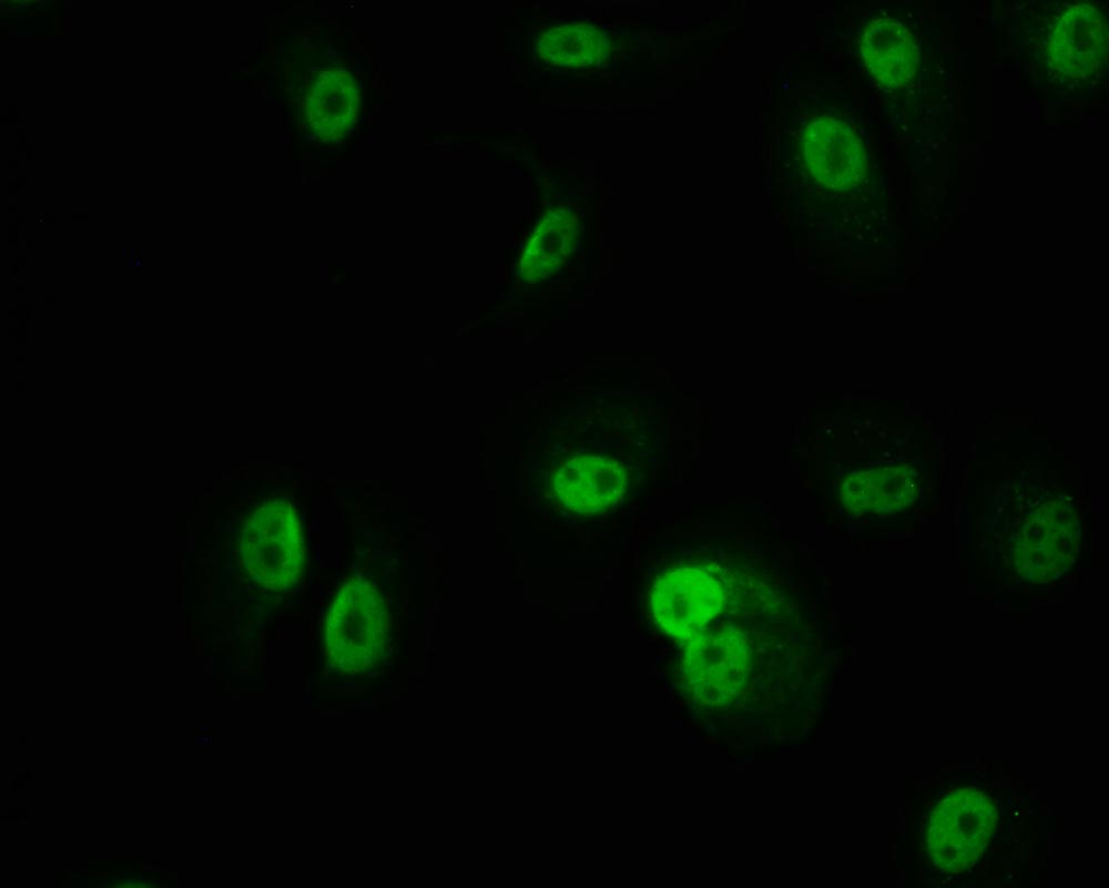
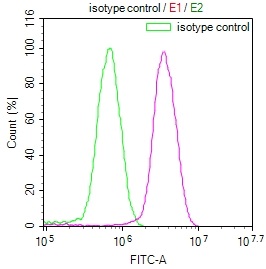
Product Details

Uniprot No.
Target Names
PKN2
Alternative Names
Serine/threonine-protein kinase N2 (EC 2.7.11.13) (PKN gamma) (Protein kinase C-like 2) (Protein-kinase C-related kinase 2), PKN2, PRK2 PRKCL2
Species Reactivity
Human
Immunogen
A synthesized peptide derived from human PKN2
Immunogen Species
Homo sapiens (Human)
Conjugate
Non-conjugated
Clonality
Monoclonal
Isotype
Rabbit IgG
Clone No.
20A8
Purification Method
Affinity-chromatography
Concentration
It differs from different batches. Please contact us to confirm it.
Buffer
Rabbit IgG in phosphate buffered saline, pH 7.4, 150mM NaCl, 0.02% sodium azide and 50% glycerol.
Form
Liquid
Tested Applications
ELISA, WB, IHC, IF, FC
Recommended Dilution
Application Recommended Dilution
WB 1:500-1:2000
IHC 1:50-1:200
IF 1:50-1:200
FC 1:50-1:200
Troubleshooting and FAQs
Storage
Upon receipt, store at -20°C or -80°C. Avoid repeated freeze.
Lead Time
Basically, we can dispatch the products out in 1-3 working days after receiving your orders. Delivery time maybe differs from different purchasing way or location, please kindly consult your local distributors for specific delivery time.
Description

The PKN2 recombinant monoclonal antibody is produced through a well-established and rigorous process to ensure its quality and specificity. Initially, B cells are isolated from an immunized animal using the synthesized peptide derived from human PKN2 as the immunogen. Total RNA is extracted from the B cells and converted into cDNA through reverse transcription. The PKN2 antibody genes are then amplified using specific primers designed for the antibody constant regions and inserted into an expression vector. This vector is transfected into host cells, enabling the production of the PKN2 recombinant monoclonal antibody. Following cell culture, the antibody is harvested from the supernatant and purified using affinity chromatography, resulting in a highly purified preparation. Extensive characterization assays, including ELISA, WB, IHC, IF, and FC analysis, are conducted to validate the antibody's specificity and functionality, ensuring its precise recognition of human PKN2 protein. This meticulous production process guarantees the generation of a reliable and effective PKN2 recombinant monoclonal antibody, suitable for diverse applications in PKN2-related research.

Customer Reviews and Q&A

 Customer Reviews

There are currently no reviews for this product.

Submit a Review here

Target Background

Function
PKC-related serine/threonine-protein kinase and Rho/Rac effector protein that participates in specific signal transduction responses in the cell. Plays a role in the regulation of cell cycle progression, actin cytoskeleton assembly, cell migration, cell adhesion, tumor cell invasion and transcription activation signaling processes. Phosphorylates CTTN in hyaluronan-induced astrocytes and hence decreases CTTN ability to associate with filamentous actin. Phosphorylates HDAC5, therefore lead to impair HDAC5 import. Direct RhoA target required for the regulation of the maturation of primordial junctions into apical junction formation in bronchial epithelial cells. Required for G2/M phases of the cell cycle progression and abscission during cytokinesis in a ECT2-dependent manner. Stimulates FYN kinase activity that is required for establishment of skin cell-cell adhesion during keratinocytes differentiation. Regulates epithelial bladder cells speed and direction of movement during cell migration and tumor cell invasion. Inhibits Akt pro-survival-induced kinase activity. Mediates Rho protein-induced transcriptional activation via the c-fos serum response factor (SRF). Involved in the negative regulation of ciliogenesis.; (Microbial infection) Phosphorylates HCV NS5B leading to stimulation of HCV RNA replication.
Gene References into Functions
  1. The expression of PKN2 in colon cancer cells suppresses tumor associated M2 macrophage polarization and tumor growth. PMID: 29368606
  2. PKN2 knockdown in vitro decreased insulin-stimulated glucose uptake, incorporation into glycogen, and oxidation. Thus, PKN2 alters key signaling pathways and transcriptional networks to regulate glucose and lipid metabolism. PMID: 28720584
  3. Helicobacter pylori CagA interacts with PRK2 and inhibits its kinase activity. PMID: 26041307
  4. findings demonstrate that Yersinia enterocolitica rYopM interacts with RSK1 and PRK2 following cell-penetration PMID: 25513777
  5. Regulation of protein kinase C-related protein kinase 2 (PRK2) by an intermolecular PRK2-PRK2 interaction mediated by Its N-terminal domain. PMID: 22511787
  6. these findings suggest that Hsp90 plays a critical role in the regulation of HCV RNA polymerase phosphorylation via the PDK1-PRK2 signaling pathway. PMID: 22490666
  7. Rho binding is essential for PRK2 function and facilitates PRK2 recruitment to junctions. Kinase-dead PRK2 acts as a dominant-negative mutant and prevents apical junction formation. PMID: 20974804
  8. PRK2/PKN2, is an essential regulator of both entry into mitosis and exit from cytokinesis in HeLa S3 cells, required for abscission of the midbody at the end of the cell division cycle and for phosphorylation and activation of Cdc25B. PMID: 17332740
  9. These observations highlight elements of Nef's functional complexity and demonstrate previously unsuspected structural requirements for PAK-2 activation and MHC-1 down-modulation in Nef's flexible N- and C-terminal regions. PMID: 17632197
  10. Little evidence of association was observed between SNPs in PKN2 and type 2 diabetes in African Americans. PMID: 18443202
  11. the last seven amino acid residues at the C-terminus of PRK2 are not required for the activation of the kinase by RhoA in vitro, however, the extreme C-terminal segment is critical for the full activation of PRK2 by RhoA in cells PMID: 18835241

Show More

Hide All

Subcellular Location
Cytoplasm. Nucleus. Membrane. Cell projection, lamellipodium. Cytoplasm, cytoskeleton. Cleavage furrow. Midbody. Cell junction.
Protein Families
Protein kinase superfamily, AGC Ser/Thr protein kinase family, PKC subfamily
Tissue Specificity
Ubiquitous. Expressed in numerous tumor cell lines, especially in bladder tumor cells.
Database Links

HGNC: 9406

OMIM: 602549

KEGG: hsa:5586

STRING: 9606.ENSP00000359552

UniGene: Hs.440833

icon of phone
Call us
301-363-4651 (Available 9 a.m. to 5 p.m. CST from Monday to Friday)
icon of address
Address
7505 Fannin St., Ste 610, Room 7 (CUBIO Innovation Center), Houston, TX 77054, USA
icon of social media
Join us with

Subscribe newsletter

Leave a message

* To protect against spam, please pass the CAPTCHA test below.
CAPTCHA verification
© 2007-2024 CUSABIO TECHNOLOGY LLC All rights reserved. 鄂ICP备15011166号-1
Place an order now

I. Product details

*
*
*
*

II. Contact details

*
*

III. Ship To

*
*
*
*
*
*
*

IV. Bill To

*
*
*
*
*
*
*
*