The Detection of Cytokines


In 1957, Alick lsaacs and Jean Lindenmann studied the interference of heat-killing influenza virus on the growth of live virus in chicken chorionic chorioallantoic membrane fragments and found that heat-inactivated influenza virus incubated with membranes produced a substance that interfered with infection and replication of the live virus[1]. This substance ( Interferon-alpha ) was the first discovered cytokine and was later named interferon because of its interfering effects.

After this, other types of interferons and other cytokines have been discovered. Scientists respectively named them according to their different functions, interleukin (IL), interferon (IFN), and tumor necrosis factor(TNF), colony stimulating factor (CSF), chemokine, growth factor (GF).


5. Comparison of Five Detection Methods


1. What are Cytokines?

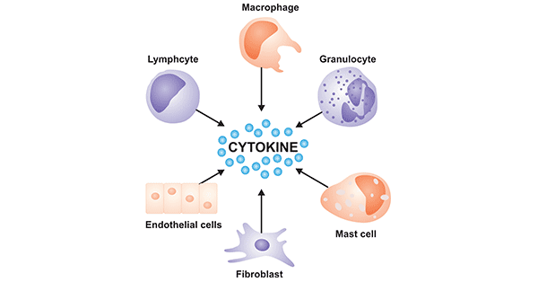
Cytokines are a class of low molecular weight polypeptide molecules that are responsible for communication between nearby cells, particularly cells of hematopoietic origin. Cytokines from cell proliferation involving inflammation, immunological, migration, fibrosis, and repair each significant biological processes angiogenesis[2][3] Cytokines produced mainly by helper T cells (Th cells) and macrophages. Although they can be transiently induced and secreted by almost all nucleated cells. A particular cytokine may be produced by more than one cell.


Various cells that produce cytokines

Figure 1  Various cells that produce cytokines

2. Role of Cytokines

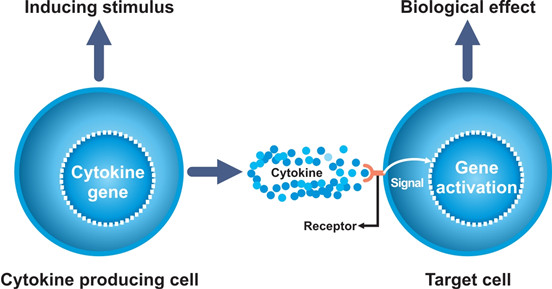
Cytokines exert biological effects by binding to corresponding cytokine receptors on the cell surface. The combination of cytokines and their receptors initiates complex intracellular molecular interactions that ultimately cause changes in cellular gene transcription.

The role of cytokines is networked. In other words, each cytokine can act on a variety of cells; each cell can be regulated by a variety of cytokines; different cytokines have synergistic or mutually restrictive effects. It thereby constitutes a complex cytokine immune regulation network.


The process by which cytokines work

Figure 2 The process by which cytokines work

3. Why Test Cytokines?

Cytokines are biomarkers of inflammation-based diseases, so almost all types of diseases involve cytokines as potential biomarkers. Elevated cytokine concentrations indicate that activation of the cytokine pathway is associated with inflammation or disease progression[4]. And studies have shown that inflammation can create an environment favorable for tumor progression[5][6].

Therefore, cytokine measurement is very important , it helps to elucidate the molecular level of immune regulation mechanism, disease prevention, diagnosis and treatment[7]. For example, interferon-γ may be useful in the treatment of chronic granulomatous disease[8] and bone sclerosis disease[9] and interferon-α for the treatment of hepatitis C[10][11] and Juvenile laryngeal papillomatosis[12]. Interferon beta is applied to treat multiple sclerosis[13], and interleukin-2 (IL-2) can treat a variety of metastatic cancer[14][15][16], in particular the use of genetic engineering techniques producing a recombinant cytokine, which has been used to treat tumors, infection, inflammation, and other hematopoietic disorders. And these therapies receive good effect and are prospective in a very broad application.


4. Cytokine Detection Methods

4.1 Real-time Quantitative Polymerase Chain Reaction Assay

Real-time quantitative polymerase chain reaction ( qPCR ) was used to study gene expression and measure cytokine mRNA transcriptional abundance. As microfluidic technology advances to improve the size and volume of picoliters[17][18], instruments that can perform PCR on a single cell can be used for single-cell studies of cytokine expression[19].

First, we should select internal reference that is not affected by the processing factor, and then use purified dsDNA, transcribed RNA and synthesized ssDNA[20]in vitro or any cDNA expressing the target gene as target genes and internal reference standards curve. Next, optimize the experimental conditions to make the two genes amplification efficiency consistent. If the efficiency is different, the calculation results will be inconsistent. Finally, the data analysis is performed using the 2-△Ct[21] method and the Ct ( cycle threshold ) value is simply used for a rough estimation.

4.2 Enzyme-linked Immunosorbent Assay

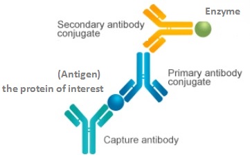
Enzyme-linked immunosorbent assay (ELISA) is a method that utilizes the specific binding of an antibody molecule to an antigen molecule, and separates the free heteroprotein from the target protein bound to the solid phase carrier, and uses a special marker to qualitatively or quantitatively analyze it. It allows the detection of cytokine secretion at the protein level. It uses an enzymatic reaction that produces color. The intensity of the color is related to the amount of protein present. ELISA platforms are widely used to quantify cytokines. When multiple cytokines in a single sample and measuring a need to develop a variety of ELISA. This is time-consuming and labor intensive and requires a relatively large number of tissue samples.

Sandwich ELISA

Figure 3 Sandwich ELISA

4.3 Enzyme-linked Immunosorbent Site Enzyme-linked Aggregate Exposure

Enzyme-linked immunosorbent site enzyme-linked aggregate exposure, also known as enzyme-linked immunospot assay (ELISPOT), is a technique for detecting proteins using antibodies. It is a modified version of an ELISA immunoassay that allows detection of cytokines from single cells[22-24]. ELISPOT is similar to ELISA because of the cytokines produced from individually sorted cells (usually Performed on a cell sorter (flow cytometer) was determined[25].

On a microplate coated with a cytokine antibody to be tested, a test cell capable of secreting a corresponding cytokine is added, and cultured in the presence or absence of a stimulant, the test cell secretes a cytokine around it, which then captured by specific antibodies on the plate. The subsequent reaction is like ELISA, after washing the cells, the enzyme-labeled antibody used for the test is a primary antibody or a secondary antibody.

One spot represents a cytokine-secreting cell, and the degree of coloration of the spot is related to the amount of cytokine secreted by the cell.

The T cell ELISpot assay can be used to characterize T cell subsets. The ELISpot assay detects cytokines IFN-γ, IL-2, TNF-α, IL-4, IL-5, and IL-13. The first three cytokines are produced by Th1 cells, while the latter three are produced by Th2 cells. Measuring T cell responses by cytokine production also makes it possible to study vaccine efficacy[26].

4.4 Cytokine Bead Array ( CBA )

The basic idea behind the bead-based capture array technology is that each type of bead has a specific protein capture antibody on its surface. Each antibody has a specific fluorescence intensity[27]. The cytokine bead array allows the use of small sample volumes to detect the entire set of cytokines[28-32] in multiple ways, namely efficiently measuring different cytokines simultaneously in a single microplate. The assay uses a flow cytometer that provides sensitivity (due to laser and optics) and velocity (fluid). Fluorescence intensity was measured by flow cytometry.

4.5 Immunohistochemistry

Immunohistochemistry (IHC) commonly used in immunostaining, by using the principle of biological tissue with an antibody that specifically binds to an antigen, tissue sections cells selectively recognize an antigen (protein)[33]. Cytokines are proteins, so this method can also be used to detect cytokines. This method can be used not only to detect the amount of antigen expressed but also to observe the position of the antigen.

The key to IHC is the choice of primary and secondary antibodies. The final fluorescence microscope can capture and interpret the image. Microscope images can be quantified and there are many "free software" programs available for this purpose, such as NIH Image and Image J, these packages will allow the determination of positive and total pixels, which can be expressed as the ratio of the measured intensity of the stain.

Of course, there are other ways to measure and determine the existence and concentration of various cytokines, such as Northern blot, reverse transcription PCR, cell or tissue in situ hybridizatio.


5.Comparison of Five Detection Methods

Detection of Cytokines Advantages Disadvantages
qPCR High sensitivity, direct, quantitative, reproducible, easy and reliable data processing, allowing detection of many different cytokines from relatively small sample volumes. Detection of relatively few samples, the presence of RNA does not always accurately reflect protein levels (like IL4, IL10 regulates post-translation), and the cell source that recognizes cytokines needs to separate different types of cells. This may be difficult, and if only a small fraction of the cells in the sample to be tested produce cytokines of interest, the threshold for detection may not be reached.
ELISA Direct, quantitative Smaller measurements (not always easy to get a sufficient amount of tissue fluid, and actual cytokine levels may be underestimated due to cytokine consumption); It is not possible to detect multiple cytokines in a single assay.
ELISPOT Low-frequency cells that produce cytokines can be quickly identified and classified. the same with ELISA
CBA Use a small sample size to detect whole cytokine panels in multiple ways; Analysis is faster and adapts to high-throughput analysis. High cost, low sensitivity, (as with ELISA) may underestimate cytokine production due to consumption.
IHC Ability to identify cell types that produce cytokines (these cells may not be found by other methods): specificity, sensitivity, simplicity, and cost. Poor quantitative, low sensitivity to detection of secreted proteins.

This article describes five methods for detecting cytokines in combination with the characteristics and functions of cytokines. Each method has its own advantages, but there are inevitably certain defects. Therefore, if you want to accurately measure the cytokine of a sample, you can combine two or more methods, because they can be compensated for each other.

In practical applications, they can be selected according to their respective experimental purposes and laboratory conditions. But no matter what method you consider to measure, the choice is based on the characteristics of the cytokine of interest.


Reference:
[1] Alick Isaacs, Jean Lindenmann. Virus interference. I. The interferon [J].   Proc. R. Soc. Lond. B Biol. Sci. 1957, 147 (927): 258–67.
[2] Finter NB, Chapman S, et al. The use of interferon-alpha in virus infections [J]. Drugs. 1991, 42(5): 749-65.
[3] Oppenheim, JJ Cytokines: past, present, and future [J].   Int. J. Hematol. 2001, 74:3-8.
[4] Dinarello CA. Proinflammatory cytokines [J]. Chest. 2000, 118: 503–8.
[5] Balkwill F, Charles KA, et al. Smoldering and polarized inflammation in the initiation and promotion of malignant disease [J]. Cancer Cell. 2005, 7:211–217.
[6] Eichten A, Coussens LM, et al. Paradoxical roles of the immune system during cancer development [J]. Nat Rev Cancer. 2006, 6:24–37.
[7] Bienvenu J, Monneret G, et al. The clinical usefulness of the measurement of cytokines.   Clinical Chemistry and Laboratory Medicine.   2000; 38: 267–85.
[8] Woodman RC, Erickson RW, et al. Prolonged recombinant interferon-gamma therapy in chronic granulomatous disease: evidence against enhanced neutrophil oxidase activity [J]. Blood. 1992, 79 (6): 1558–62.
[9] Key LL, Rodriguiz RM, et al. Long-term treatment of osteopetrosis with recombinant human interferon gamma [J].  N. Engl. J. Med. 1995, 332 (24): 1594–9.
[10] Finter NB, Chapman S, et al. The use of interferon-alpha in virus infections [J]. Drugs. 1991, 42 (5): 749-65.
[11] Hoofnagle JH, Seeff LB. Peginterferon and ribavirin for chronic hepatitis C [J]. N Engl J Med. 2006, 355: 2444–51.
[12] Denuas L, Alcantud V, et al. Use of interferon-alpha in laryngeal papillomatosis: eight years of a Cuban national programme [J]. J Laryngol Otol. 1997, 111: 134–40.
[13] Jacobs L, O'Malley J, et al. Intrathecal interferon in multiple sclerosis [J]. Arch Neurol. 1982, 39:609–15.
[14] Rosenberg SA, JC Yang, et al. Treatment of 283 consecutive patients with metastatic melanoma or renal cell cancer using high-dose bolus interleukin 2 [J]. JAMA. 1994, 271: 907–913.
[15] Rosenberg   SA,   JC   Yang, et al. Durability of complete responses in patients with metastatic cancer treated with high-dose interleukin-2: identification of the antigens mediating response [J].  Ann. Surg. 1998, 228: 307–319.
[16]   Yang   JC,   RM   Sherry, et al. Randomized study of high-dose and low-dose interleukin-2 in patients with metastatic renal cancer [J].  J. Clin. Oncol. 2003, 21:  3127–3132.
[17] Marcus JS, Anderson WF, et al. Microfluidic Single-Cell mRNA Isolation and Analysis [J]. Anal Chem. 2006, 78:3084–9.
[18] Marcus JS, Anderson WF, et al. Parallel Picoliter RT-PCR Assays Using Microfluidics [J].   Anal Chem.   2006;78:956–8.
[19] Diercks A, Kostner H, et al. Resolving cell population heterogeneity: real-time PCR for simultaneous multiplexed gene detection in multiple single-cell samples [J]. PLoS One. 2009, 4   No pp. given.
[20] Bustin SA. Absolute quantification of mRNA using real-time reverse transcription polymerase chain reaction assays [J]. J Mol Endocrinol. 2000, 25:169-193.
[21] Allan SM, Harrison DC, et al. Selective increases in cytokine expression in the rat brain in response to striatal injection of a-amino-3-hydroxy-5-methyl-4-isoxazolepropionate and interleukin-1 [J]. Molecul Brain Res. 2001, 93:180-189.
[22] Favre N, Bordmann G, et al. Comparison of cytokine measurements using ELISA, ELISPOT and semi-quantitative RT-PCR [J].  J Immunol Methods. 1997, 204: 57–66.
[23] Ekerfelt C, Ernerudh J, et al. Detection of spontaneous and antigen-induced human interleukin-4 responses in vitro: comparison of ELISPOT, a novel ELISA and real-time RT-PCR [J].   J Immunol Methods. 2002, 260: 55–67.
[24] Atochina O, Mylvaganam R, et al. Comparison of results using the gel microdrop cytokine secretion assay with ELISPOT and intracellular cytokine staining assay [J].  Cytokine. 2004, 27:120–8.
[25] Kalyuzhny AE.  Methods in Molecular Biology. Totowa, NJ: Humana Press; 2012. Handbook of ELISPOT: Methods and Protocols.
[26] Strand, Nacka. Find 1 cell in 100,000 with ELISpot. MABTECH. Retrieved   6 December   2018.
[27] Elshal MF and McCoy JP. Multiplex Bead Array Assays: Performance Evaluation and Comparison of Sensitivity to ELISA [J]. Methods. 2006, 38: 317–23.
[28] Carson RT, Vignali DAA. Simultaneous quantitation of 15 cytokines using a multiplexed flow cytometric assay [J]. J Immunol Methods. 1999, 227:41–52.
[29] Earley MC, Vogt RF, et al. Report from a workshop on multianalyte microsphere assays [J].   Cytometry. 2002, 50:239–42.
[30] Vignali DA. Multiplexed particle-based flow cytometric assays. J Immunol Methods[J]. 2000, 243:243–55.
[31] Khan SS, Smith MS, et al. Multiplex bead array for detection of soluble cytokines: comparisons of sensitivity and quantitative values among kits from multiple manufacturers [J].   Cytometry, Part B. 2004, 61B: 35–9.
[32] Morgan E, Varro R, et al. Cytometric bead array: a multiplexed assay platform with applications in various areas of biology [J].   Clin Immunol. 2004, 110: 252–66.


Cite this article

CUSABIO team. The Detection of Cytokines. https://www.cusabio.com/c-20931.html
 

Comments

Leave a Comment

Your email address will not be published. Required fields are marked *
*
*
*CAPTCHA verification
icon of phone
Call us
301-363-4651 (Available 9 a.m. to 5 p.m. CST from Monday to Friday)
icon of address
Address
7505 Fannin St., Ste 610, Room 7 (CUBIO Innovation Center), Houston, TX 77054, USA
icon of social media
Join us with

Subscribe newsletter

Leave a message

* To protect against spam, please pass the CAPTCHA test below.
CAPTCHA verification
© 2007-2024 CUSABIO TECHNOLOGY LLC All rights reserved. 鄂ICP备15011166号-1