Call us
301-363-4651 (Available 9 a.m. to 5 p.m. CST from Monday to Friday)
LYPD3 (Ly6/PLAUR Domain-Containing Protein 3), also known as C4.4A, is a glycosylphosphatidylinositol (GPI)-anchored cell membrane protein that plays a key role in tumor progression, cell migration, and tissue repair. In recent years, LYPD3 has garnered significant attention in the field of tumor biomedicine. Studies have shown that it exhibits significantly high expression in various malignant tumors, including breast cancer, non-small cell lung cancer (NSCLC), acute myeloid leukemia (AML), and melanoma.
Numerous studies have confirmed that LYPD3 expression is significantly higher in various tumor tissues compared to normal tissues. In breast cancer, LYPD3 expression is regulated by β2-adrenergic signaling, promoting tumor cell migration and invasion; knocking down LYPD3 significantly inhibits tumor cell activity. Notably, LYPD3 is expressed in both primary and metastatic breast cancer lesions but is undetectable in normal breast tissue [1]. Furthermore, in lung adenocarcinoma (LUAD), LYPD3 expression is significantly upregulated in tumor tissue compared to adjacent non-tumor tissue. More critically, high LYPD3 expression shows significant associations with patient ethnicity, tumor stage, and survival status. Specifically, higher LYPD3 expression levels correlate with poorer patient survival, and LYPD3 expression levels significantly increase with advancing stages of LUAD [2].
The critical role of LYPD3 in tumor invasion and metastasis has been confirmed by multiple studies. In NSCLC, high LYPD3 expression is associated with tumor cell proliferation, inhibition of apoptosis, and an invasive tumor phenotype[2]. Simultaneously, in head and neck squamous cell carcinoma and non-small cell lung cancer, LYPD3 interacts with miRNAs (such as miR-151-5p, miR-124-3p) and long non-coding RNAs (such as OGFRP1) to regulate tumor cell migration, invasion, and proliferation. Its high expression is closely associated with poor patient prognosis [3][4]. Research also indicates that LYPD3 promotes tumor cell self-renewal and drug resistance by maintaining cancer stemness properties, thereby exacerbating tumor invasiveness and metastatic potential. This has been demonstrated in various tumors including melanoma and colorectal cancer [5][6].
LYPD3 plays an important role in shaping the tumor immune microenvironment. In melanoma, LYPD3 is co-expressed with the immunosuppressive marker S100A9 and is associated with an immunosuppressive tumor microenvironment. Its expression negatively correlates with the number of tumor-infiltrating T cells, suggesting that LYPD3 may influence tumor development and treatment response by regulating immune cell infiltration [5]. Additionally, in AML, high LYPD3 expression is associated with poor prognosis and may be involved in AML development by regulating the P53 and/or PI3K/AKT signaling pathways. Experimental data show that inhibiting LYPD3 expression using siRNA suppresses the proliferation of the AML cell line HL-60 and significantly upregulates apoptosis-related markers, indicating that LYPD3 plays a crucial role in promoting the proliferation and survival of AML cells [7].
Given the aberrant expression of LYPD3 in various tumors and its close relationship with tumor progression, LYPD3 holds promise as a novel therapeutic target for cancer treatment. In breast cancer, LYPD3 expression, regulated by β2-adrenergic signaling, promotes tumor cell migration and invasion. Knocking down LYPD3 significantly inhibits tumor cell activity, highlighting its potential as a therapeutic target [1]. In melanoma, the JUP/AGR2/LYPD3 signaling axis not only maintains cancer stemness but also promotes glycolysis and is closely linked to an immunosuppressive tumor microenvironment, suggesting LYPD3 contributes to tumor immune escape [5]. Furthermore, in lung cancer, high LYPD3 expression is closely associated with poor prognosis, tumor stage, and immune cell infiltration, and affects multiple signaling pathways including the cell cycle and DNA replication [8]. Importantly, LYPD3 is nearly absent in normal tissues except for skin keratinocytes and esophageal dermal cells. This characteristic gives LYPD3-targeted therapy high specificity, promising precise targeting of tumor cells while minimizing damage to normal tissues. Synthesizing the multiple key mechanisms of LYPD3 in tumor regulation, it has emerged as an important molecule in tumor diagnosis, prognosis assessment, and targeted therapy, holding the potential to open new avenues for solid tumor treatment and offer patients more effective therapeutic options and better prognoses.
The main type of investigational drugs targeting LYPD3 is monoclonal antibodies, indicated for tumors. The following is the pipeline of LYPD3-targeting monoclonal antibodies in development:
| Drug | Drug Type | Indication | Developers | Highest Phase |
|---|---|---|---|---|
| GT-002 (PentixaPharm) | Monoclonal Antibody | Tumor | PentixaPharm GmbH | Preclinical |
| GT-002 (Glycotope) | Monoclonal Antibody | Tumor | Glycotope GmbH | Drug Discovery |
(Data source: Pharmsnap)
● LYPD3 Recombinant Proteins
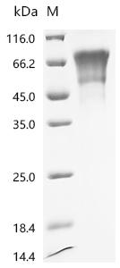
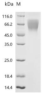
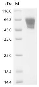
Recombinant Human Ly6/PLAUR domain-containing protein 3 (LYPD3) (Active); CSB-MP013263HU

Recombinant Mouse Ly6/PLAUR domain-containing protein 3 (Lypd3), partial (Active); CSB-MP849681MO1

Recombinant Rat Ly6/PLAUR domain-containing protein 3 (Lypd3) (Active); CSB-MP013263RA
● LYPD3 Recombinant Antibody
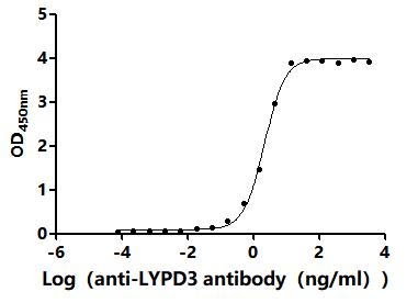
LYPD3 (Lupartumab) Recombinant Monoclonal Antibody; CSB-RA013263MA2HU
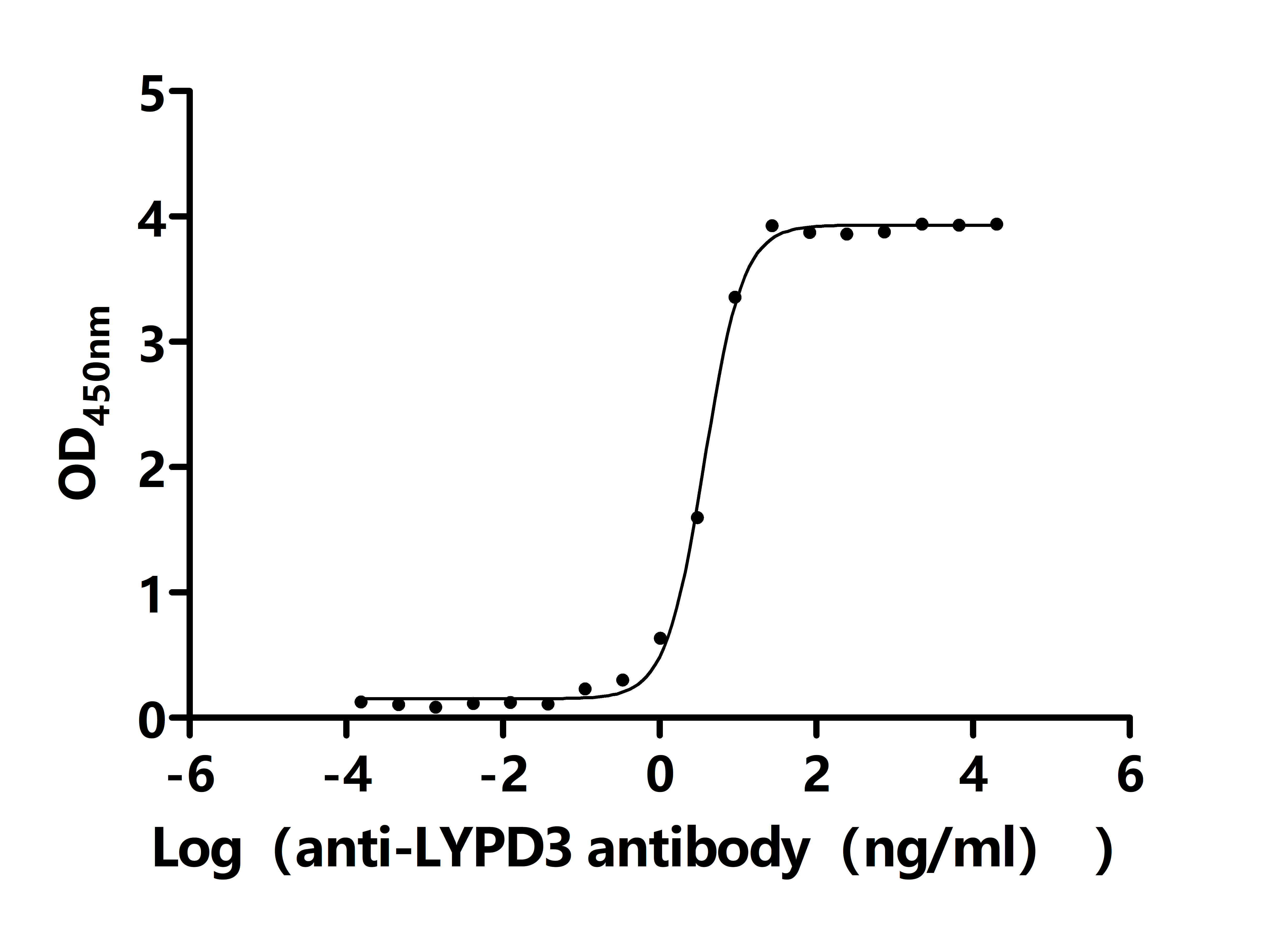
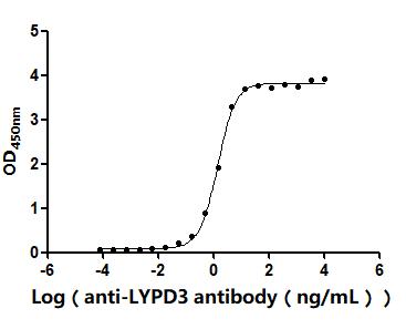
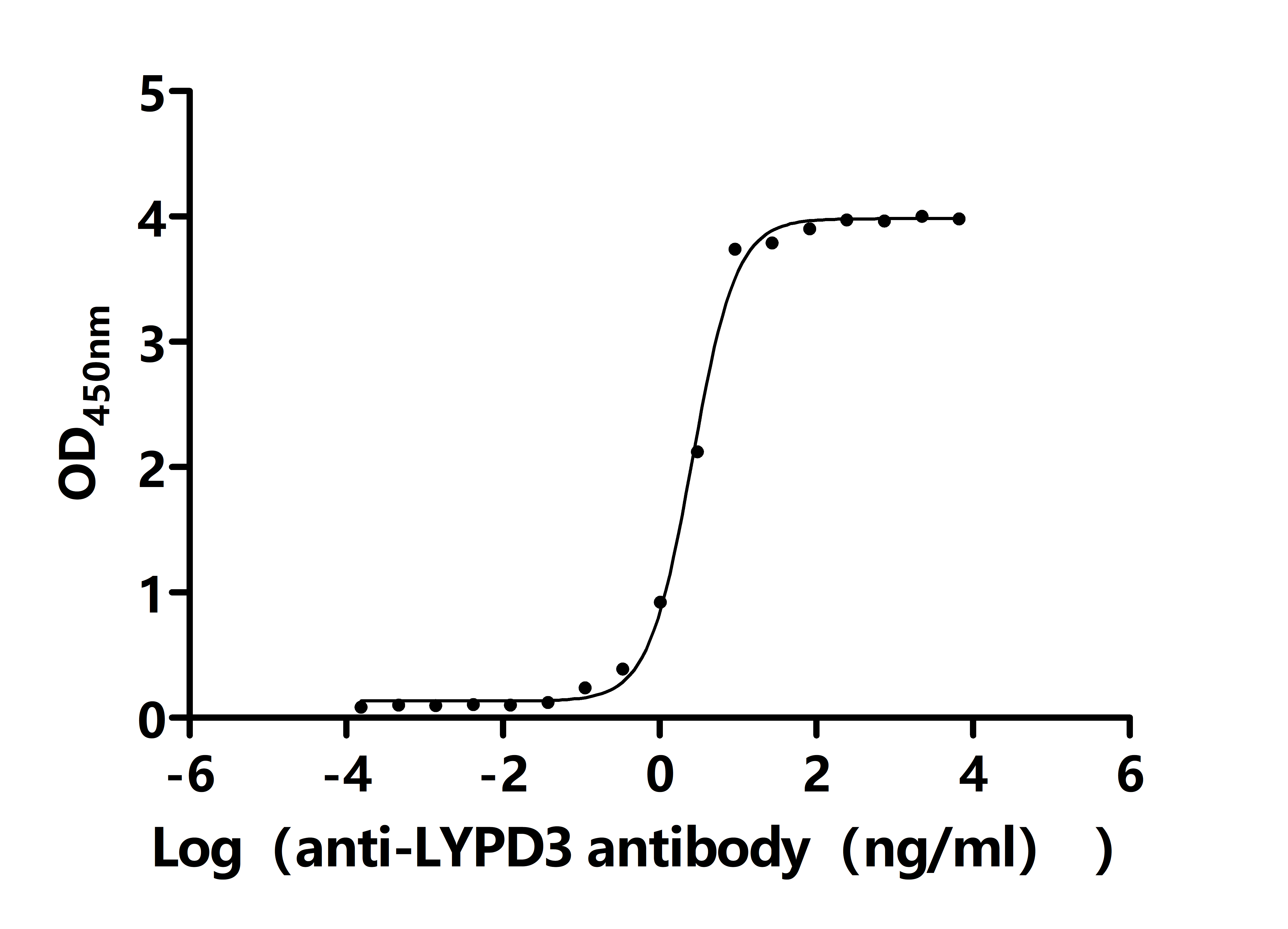
The Binding Activity of Human LYPD3 with Anti-LYPD3 recombinant antibody
Activity: Measured by its binding ability in a functional ELISA. Immobilized Human LYPD3(CSB-MP013263HU) at 2 μg/mL can bind Anti-LYPD3 recombinant antibody. The EC50 is 2.375-2.905 ng/mL.
References
[1] Gruet, M., Cotton, D., Coveney, C., Boocock, D., Wagner, S., Komorowski, L., Rees, R., Pockley, A., Garner, A., Wallis, J., Miles, A., Powe, D., & Powe, D. (2020). β2-Adrenergic Signalling Promotes Cell Migration by Upregulating Expression of the Metastasis-Associated Molecule LYPD3. Biology, 9.
[2] Ying Huang et al. "Elevated Expression of LYPD3 Is Associated with Lung Adenocarcinoma Carcinogenesis and Poor Prognosis.." DNA and cell biology (2020).
[3] Yufei Hua et al. "m6A-dependent mature miR-151-5p accelerates the malignant process of HNSCC by targeting LYPD3." Molecular Biomedicine, 5 (2024).
[4] Yan Zhang et al. "Long non-coding RNA OGFRP1 regulates LYPD3 expression by sponging miR-124-3p and promotes non-small cell lung cancer progression.." Biochemical and biophysical research communications, 505 2 (2018): 578-585.
[5] Yuan-Jie Liu et al. "LY6/PLAUR domain containing 3 (LYPD3) maintains melanoma cell stemness and mediates an immunosuppressive microenvironment." Biology Direct, 18 (2023).
[6] T. Kanaseki et al. "LY6/PLAUR domain containing 3 has a role in the maintenance of colorectal cancer stem-like cells.." Biochemical and biophysical research communications, 486 2 (2017): 232-238.
[7] Tingting Hu et al. "LYPD3, a New Biomarker and Therapeutic Target for Acute Myelogenous Leukemia." Frontiers in Genetics, 13 (2022).
[8] Xinyao Xu et al. "Comprehensive analysis of exosome gene LYPD3 and prognosis/immune cell infiltration in lung cancer." Translational Cancer Research, 13 (2023): 1394 - 1405.


Comments
Leave a Comment