Call us
301-363-4651 (Available 9 a.m. to 5 p.m. CST from Monday to Friday)
| Code | CSB-MP004952HU |
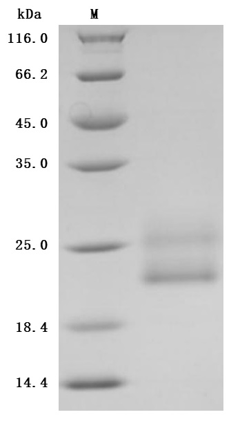
| Abbreviation | Recombinant Human CD69 protein, partial (Active) |
| MSDS | |
| Size | $9.9 |
| Promotion |

|
| Order now | |
| Image |
|
| Have Questions? | Leave a Message or Start an on-line Chat |
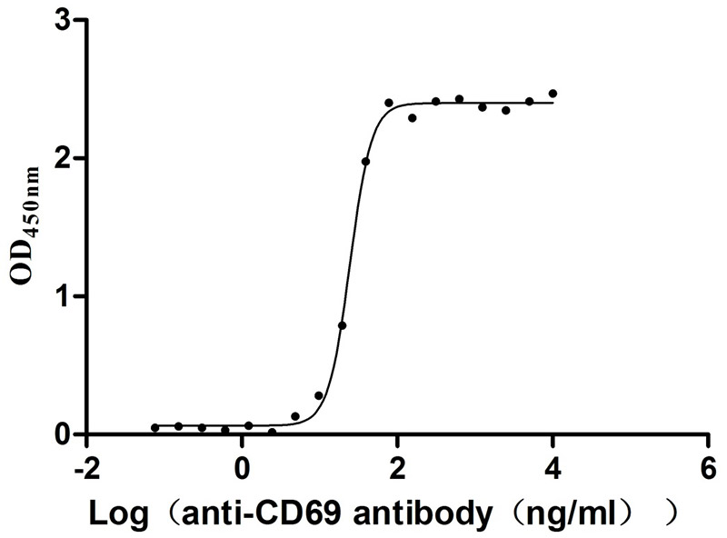
CD69 functions as an early lymphocyte activation marker and regulator of immune cell egress from lymphoid tissues, making it a critical target for understanding T-cell activation dynamics and inflammatory responses. This mammalian-expressed construct spanning residues 62–199 captures the extracellular C-type lectin domain responsible for receptor function, and the expression system ensures native glycosylation patterns essential for proper folding and antibody recognition. Functional ELISA validation demonstrates specific antibody binding with an EC50 of 23.17–26.04 ng/mL, confirming conformational integrity suitable for therapeutic antibody epitope mapping, competitive inhibition screening, and ligand-receptor interaction studies by ELISA or surface plasmon resonance. The protein meets quality thresholds commonly required for antibody validation workflows, with purity exceeding 95% and endotoxin levels below 1.0 EU/μg, providing a reliable positive control for binding assays in cancer immunology and immune checkpoint research.
There are currently no reviews for this product.

