Recombinant Human Glial fibrillary acidic protein (GFAP)

In Stock
Code CSB-EP009369HUa0
Abbreviation Recombinant Human GFAP protein
MSDS
Size $256
Order now
Image
  • (Tris-Glycine gel) Discontinuous SDS-PAGE (reduced) with 5% enrichment gel and 15% separation gel.
Have Questions? Leave a Message or Start an on-line Chat

Product Details

Purity
Greater than 90% as determined by SDS-PAGE.
Target Names
Uniprot No.
Research Area
Neuroscience
Species
Homo sapiens (Human)
Source
E.coli
Expression Region
1-432aa
Target Protein Sequence
MERRRITSAARRSYVSSGEMMVGGLAPGRRLGPGTRLSLARMPPPLPTRVDFSLAGALNAGFKETRASERAEMMELNDRFASYIEKVRFLEQQNKALAAELNQLRAKEPTKLADVYQAELRELRLRLDQLTANSARLEVERDNLAQDLATVRQKLQDETNLRLEAENNLAAYRQEADEATLARLDLERKIESLEEEIRFLRKIHEEEVRELQEQLARQQVHVELDVAKPDLTAALKEIRTQYEAMASSNMHEAEEWYRSKFADLTDAAARNAELLRQAKHEANDYRRQLQSLTCDLESLRGTNESLERQMREQEERHVREAASYQEALARLEEEGQSLKDEMARHLQEYQDLLNVKLALDIEIATYRKLLEGEENRITIPVQTFSNLQIRETSLDTKSVSEGHLKRNIVVKTVEMRDGEVIKESKQEHKDVM
Note: The complete sequence may include tag sequence, target protein sequence, linker sequence and extra sequence that is translated with the protein sequence for the purpose(s) of secretion, stability, solubility, etc.
If the exact amino acid sequence of this recombinant protein is critical to your application, please explicitly request the full and complete sequence of this protein before ordering.
Mol. Weight
54.0 kDa
Protein Length
Full Length
Tag Info
N-terminal 6xHis-tagged
Form
Liquid or Lyophilized powder
Note: We will preferentially ship the format that we have in stock, however, if you have any special requirement for the format, please remark your requirement when placing the order, we will prepare according to your demand.
Buffer
If the delivery form is liquid, the default storage buffer is Tris/PBS-based buffer, 5%-50% glycerol. If the delivery form is lyophilized powder, the buffer before lyophilization is Tris/PBS-based buffer, 6% Trehalose.
Reconstitution
We recommend that this vial be briefly centrifuged prior to opening to bring the contents to the bottom. Please reconstitute protein in deionized sterile water to a concentration of 0.1-1.0 mg/mL.We recommend to add 5-50% of glycerol (final concentration) and aliquot for long-term storage at -20°C/-80°C. Our default final concentration of glycerol is 50%. Customers could use it as reference.
Troubleshooting and FAQs
Storage Condition
Store at -20°C/-80°C upon receipt, aliquoting is necessary for mutiple use. Avoid repeated freeze-thaw cycles.
Shelf Life
The shelf life is related to many factors, storage state, buffer ingredients, storage temperature and the stability of the protein itself.
Generally, the shelf life of liquid form is 6 months at -20°C/-80°C. The shelf life of lyophilized form is 12 months at -20°C/-80°C.
Lead Time
3-7 business days
Notes
Repeated freezing and thawing is not recommended. Store working aliquots at 4°C for up to one week.
Datasheet & COA
Please contact us to get it.
Description

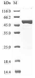
Recombinant Human Glial fibrillary acidic protein (GFAP) is produced using an E.coli expression system and contains the complete protein sequence from amino acids 1 to 432. The protein carries an N-terminal 6xHis-tag that simplifies purification and detection processes. SDS-PAGE analysis confirms a purity level exceeding 90%, which appears to provide reliable quality for research applications. This product is intended for research use only and is not for diagnostic or therapeutic purposes.

GFAP is a key intermediate filament protein that's predominantly found in astrocytes within the central nervous system. It plays what seems to be a crucial role in maintaining the structural integrity and function of neural cells. The protein is involved in various cellular processes, including cell communication and the modulation of synaptic activity. This makes it particularly significant for studies focused on neurodegenerative diseases and neural development.

Potential Applications

Note: The applications listed below are based on what we know about this protein's biological functions, published research, and experience from experts in the field. However, we haven't fully tested all of these applications ourselves yet. We'd recommend running some preliminary tests first to make sure they work for your specific research goals.

Based on the provided information, the recombinant Human Glial Fibrillary Acidic Protein (GFAP) is expressed in E. coli, a prokaryotic system that is generally unsuitable for producing properly folded eukaryotic intermediate filament proteins. GFAP requires specific folding, assembly into complex filament structures, and may need post-translational modifications that E. coli cannot provide. The presence of an N-terminal 6xHis tag may further interfere with proper folding and filament formation. While the protein is full-length (1-432aa) with >90% purity, the E. coli expression system significantly increases the risk of misfolding. Since activity is explicitly unverified, the protein cannot be assumed to be correctly folded or bioactive without experimental validation.

1. Antibody Development and Validation

This application is appropriate. The recombinant GFAP can serve as an effective immunogen for antibody generation, as antibodies may recognize linear epitopes even if the protein is misfolded. The His tag facilitates purification and screening. However, antibodies may not recognize conformational epitopes of native GFAP in astrocytes if the protein is misfolded. Validation against native GFAP from mammalian sources is recommended.

2. Protein-Protein Interaction Studies

This application requires caution. The His tag enables technical feasibility for pull-down assays, but if GFAP is misfolded, interactions may not be physiological. The protein may not properly interact with authentic binding partners in intermediate filament networks. This application should only be pursued after confirming proper folding through biophysical characterization.

3. Biochemical Characterization and Stability Studies

This application is well-suited and should be prioritized. Techniques like circular dichroism spectroscopy, dynamic light scattering, and thermal stability assays can directly assess the protein's folding state and stability. These studies are valuable even if the protein is misfolded, as they characterize the recombinant product itself.

4. In Vitro Filament Assembly Assays

This application is highly dependent on correct folding. If GFAP is misfolded, it will not assemble into proper intermediate filaments, leading to misleading results about assembly mechanisms. This application requires prior validation of proper folding and assembly capability through electron microscopy or other structural analyses.

Final Recommendation & Action Plan

Given the high probability of misfolding in E. coli for this complex eukaryotic protein, we recommend first performing a comprehensive biophysical characterization (e.g., circular dichroism for secondary structure, size-exclusion chromatography for oligomeric state) to assess folding quality. Antibody development can proceed immediately, while protein interaction and filament assembly studies should be deferred until proper folding is validated. For filament assembly assays, include positive controls with native GFAP when possible. If misfolding is detected, focus applications on biochemical characterization and antibody development rather than functional studies. Consider using eukaryotic expression systems for research requiring properly folded GFAP with native functionality.

Customer Reviews and Q&A

 Customer Reviews

There are currently no reviews for this product.

Submit a Review here

Target Background

Function
GFAP, a class-III intermediate filament, is a cell-specific marker that, during the development of the central nervous system, distinguishes astrocytes from other glial cells.
Gene References into Functions
  1. The features of the neuropathology and immunopathology of GFAP astrocytopathies were perivascular inflammation and loss of astrocytes and neurons PMID: 29193473
  2. amniotic fluid -GFAP levels differentiate between myelomeningocele and myeloschisis, and raise interesting questions regarding the clinical significance between the 2 types of defects. PMID: 28768252
  3. Desmin, Glial Fibrillary Acidic Protein, Vimentin, and Peripherin are type III intermediate filaments that have roles in health and disease [review] PMID: 29196434
  4. Plasma concentration of GFAP demonstrated associations with stroke occurrence in a West African cohort but was not associated with stroke severity or mortality. PMID: 29074065
  5. This study demonstrated that Concentrations of microparticles expressing GFAP and AQP4 were significantly higher in the traumatic brain injury group compared with healthy controls. PMID: 28972406
  6. The authors observed higher serum levels of GFAP and UCH-L1 in brain-injured children compared with controls and also demonstrated a step-wise increase of biomarker concentrations over the continuum of severity from mild to severe traumatic brain injury. Serum UCH-L1 and GFAP concentrations also strongly predicted poor outcome. PMID: 27319802
  7. Study examined if QKI6B expression can predict the outcome of GFAP, and several oligodendrocyte-related genes, in the prefrontal cortex of brain samples of schizophrenic individuals. QKI6B significantly predicts the expression of GFAP, but does not predict oligodendrocyte-related gene outcome, as previously seen with other QKI isoforms. PMID: 28552414
  8. GFAP, along with tau and AmyloidBeta42, were increased in plasma up to 90 days after traumatic brain injury compared with controls. PMID: 27312416
  9. Results show that the positive rates and expression levels of nestin, tyrosine hydroxylase (TH), GFAP and IL-17 were significantly decreased while Foxp3 and the ratio of Foxp3/IL-17 were statistically elevated in BM of AML patients. PMID: 27016413
  10. GFAP levels >0.29 ng/ml were seen only in intracerebral hemorrhage, thus confirming the diagnosis of ICH during prehospital care. PMID: 27951536
  11. These results indicate that autoantibodies against GFAP could serve as a predictive marker for the development of overt autoimmune diabetes. PMID: 28546444
  12. Higher median plasma GFAP values were documented in intracerebral hemorrhage compared with acute ischemic stroke, stroke mimics, and controls. PMID: 28751552
  13. GFAP is specifically expressed in the auricular chondrocytes, and assumes a pivotal role in resistance against mechanical stress. PMID: 28063220
  14. Bevacizumab treatment was also associated with structural protein abnormalities, with decreased GFAP and vimentin content and upregulated GFAP and vimentin mRNA expression. PMID: 28419863
  15. the exchange of GFP-GFAPdelta was significantly slower than the exchange of GFP-GFAPalpha with the intermediate filament-network. PMID: 27141937
  16. Tat expression or GFAP expression led to formation of GFAP aggregates and induction of unfolded protein response (UPR) and endoplasmic reticulum (ER) stress in astrocytes. PMID: 27609520
  17. This study demonstrated that GFAP exhibited distinct temporal profiles over the course of 7 days in patient with traumatic brain injury. PMID: 27018834
  18. e data indicates that serum GFAP levels may be associated with severity of autism spectrum disorders among Chinese children. PMID: 28088366
  19. High GFAP expression is associated with retinoblastoma. PMID: 27488116
  20. Overall, glial fibrillary acidic protein reflected no evidence for significant peripartum brain injury in neonates with congenital heart defects, but there was a trend for elevation by postnatal day 4 in neonates with left heart obstruction. PMID: 26786018
  21. serum levels of GFAP were significantly lower in autism spectrum disorders than controls PMID: 27097671
  22. We found downregulation of GFAP mRNA and protein in the mediodorsal thalamus and caudate nucleus of depressed suicides compared with controls, whereas GFAP expression in other brain regions was similar between groups. Furthermore, a regional comparison including all samples revealed that GFAP expression in both subcortical regions was, on average, between 11- and 15-fold greater than in cerebellum and neocortex. PMID: 26033239
  23. No difference in cord blood concentration found between hypoxic-ischemic encephalopathy neonates and controls PMID: 26135781
  24. GFAP is upregulated following an insult or injury to the brain, additionally making it an indicator of CNS pathology. PMID: 25846779
  25. This study demonistrated that the density of GFAP-immunoreactive astrocytes is decreased in left hippocampi in major depressive disorder PMID: 26742791
  26. This study demonstrated that GFAP as a promising biomarker to distinguish ischemic stroke from intracerebral hemorrhage. PMID: 26526443
  27. The levels of GFAP in Alzheimer's disease, dementia with Lewy bodies, and frontotemporal lobar degeneration patients were significantly higher than those in the healthy control subjects. PMID: 26485083
  28. GFAP is significantly associated with outcome, but it does not add predictive power to commonly used prognostic variables in a population of patients with TBI of varying severities. PMID: 26547005
  29. Neither duplications nor deletions of GFAP were found, suggesting that GFAP coding-region rearrangements may not be involved in Alexander disease or Alexanderrelated leukoencephalopathies. PMID: 26208460
  30. The data suggest that human vitreous body GFAP is a protein biomarker for glial activation in response to retinal pathologies. PMID: 26279003
  31. Studied diagnostic Value of Serum Levels of GFAP, pNF-H, and NSE Compared With Clinical Findings in Severity Assessment of Human Traumatic Spinal Cord Injury. PMID: 25341992
  32. GFAP peaks early during haemorrhagic brain lesions (at significantly higher levels), and late in ischaemic events, whereas antibodies against NR2 RNMDA have significantly higher levels during ischemic stroke at all time-points. PMID: 26081945
  33. There was an absence of GFAP in astrocytes during early fetal spinal cord development until 9 months of gestation , and the appearance of GFAP-positive reactivity was later than that of neurons. PMID: 25904356
  34. It could be a clinically relevant marker associated with tumor invasiveness in cerebral astrocytomas. PMID: 25178519
  35. These data imply that a tight regulation of histone acetylation in astrocytes is essential, because dysregulation of gene expression causes the aggregation of GFAP, a hallmark of human diseases like Alexander's disease. PMID: 25128567
  36. Identification of a novel nonsense mutation in the rod domain of GFAP that is associated with Alexander disease. PMID: 24755947
  37. The role of S100B protein, neuron-specific enolase, and glial fibrillary acidic protein in the evaluation of hypoxic brain injury in acute carbon monoxide poisoning PMID: 24505052
  38. GFAP, the principal intermediate filament protein of astrocytes, is involved in physiological, but in particular, in pathophysiological functions of astrocytes, the latter ones being connected with astrocyte activation and reactive gliosis. [Review] PMID: 25726916
  39. The data on the changes in expression of GFAP in Alexander disease caused by the primary pathology of astrocytes are presented. PMID: 25859599
  40. A combined profile of preoperative IGFBP-2, GFAP, and YKL-40 plasma levels could serve as an additional diagnostic tool for patients with inoperable brain lesions suggestive of Glioblastoma multiforme. PMID: 25139333
  41. There are significant increases in glial fibrillary acidic protein levels in children undergoing cardiopulmonary bypass for repair of congenital heart disease. The highest values were seen during the re-warming phase. PMID: 23845562
  42. This stuidy demonistrated that Fibrillary astrocytes are decreased in the subgenual cingulate in schizophrenia. PMID: 24374936
  43. TBI patients showed an average 3.77 fold increase in anti-GFAP autoantibody levels from early (0-1 days) to late (7-10 days) times post injury. PMID: 24667434
  44. We showed that GFAP is over-expressed and hypophosphorylated in the enteric glial cells of Parkinson's disease patients as compared to healthy subjects PMID: 24749759
  45. Its expression is associated with plaque load related astrogliosis in Alzheimer's disease. PMID: 24269023
  46. The findings of this study that caspase-mediated GFAP proteolysis may be a common event in the context of both the GFAP mutation and excess. PMID: 24102621
  47. This study demonistratedt hat Increased expression of glial fibrillary acidic protein in prefrontal cortex in psychotic illness PMID: 23911257
  48. Data indicate that Gfapdelta is expressed in the in developing mouse brain sub-ventricular zones in accordance with the described localization in the developing and adult human brain. PMID: 23991052
  49. GFAP-breakdown products blood levels reliably distinguished severity of injury in traumatic brain injury patients. PMID: 23489259
  50. The C/C genotype at rs2070935 of the GFAP promoter in late-onset AxD was associated with an earlier onset and a more rapid progression of ambulatory disability compared with the other genotypes. PMID: 23903069

Show More

Hide All

Involvement in disease
Alexander disease (ALXDRD)
Subcellular Location
Cytoplasm.
Protein Families
Intermediate filament family
Tissue Specificity
Expressed in cells lacking fibronectin.
Database Links

HGNC: 4235

OMIM: 137780

KEGG: hsa:2670

STRING: 9606.ENSP00000253408

UniGene: Hs.514227

icon of phone
Call us
301-363-4651 (Available 9 a.m. to 5 p.m. CST from Monday to Friday)
icon of address
Address
7505 Fannin St., Ste 610, Room 7 (CUBIO Innovation Center), Houston, TX 77054, USA
icon of social media
Join us with

Subscribe newsletter

Leave a message

* To protect against spam, please pass the CAPTCHA test below.
CAPTCHA verification
© 2007-2025 CUSABIO TECHNOLOGY LLC All rights reserved. 鄂ICP备15011166号-1
×
Place an order now

I. Product details

*
*
*
*

II. Contact details

*
*

III. Ship To

*
*
*
*
*
*
*

IV. Bill To

*
*
*
*
*
*
*
*