Call us
301-363-4651 (Available 9 a.m. to 5 p.m. CST from Monday to Friday)
Antibodies are indispensable tools for biomedical research, powering core techniques like Western blot (WB), immunoprecipitation (IP), immunohistochemistry (IHC), immunofluorescence (IF), chromatin immunoprecipitation (ChIP), and flow cytometry (FC). Yet up to 50% of commercially available antibodies fail to meet basic performance standards [1,2], leading to wasted time, reagents, and funding—costing the U.S. research community an estimated $350 million to $1.8 billion annually [1]. If your experiment yields inconsistent or unexpected results, the first question to ask is: Did I choose the right antibody?
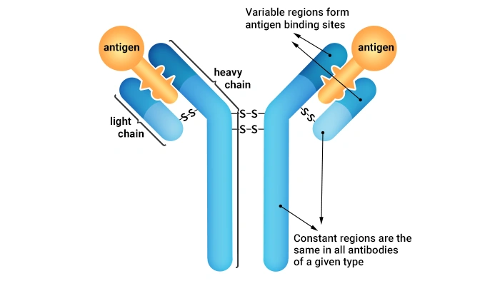
Figure 1. Antibody structure (Y-shaped type)
Even experienced researchers struggle with antibody selection. For understudied proteins or rare model systems, options may be limited. For well-characterized targets, the overwhelming number of products from hundreds of vendors creates a different challenge. This guide simplifies the process with practical, evidence-based recommendations to help you select high-performing antibodies for your experiments.
Table of Contents
Specificity is the most critical attribute of a research antibody. It determines whether your antibody binds only to the target protein and not to unrelated molecules. When evaluating specificity, consider four key dimensions:
Each antibody binds to a unique epitope (a 5–15 amino acid sequence) on the target protein (Figure 1). To ensure your antibody recognizes your specific target:
Most commercial antibodies are raised against human or mouse antigens. While many cross-react with other species due to sequence homology, never assume this is the case:
Antibody performance is highly application-dependent because sample preparation methods alter protein conformation [6]
Never use an antibody for an application it has not been validated for unless you are prepared to perform extensive validation yourself.
Different techniques have unique requirements for antibody performance. Use these guidelines to select the right antibody for your application:
WB detects denatured, linearized proteins separated by gel electrophoresis.
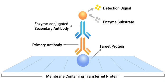
Figure 2. WB Principle Diagram
IP and ChIP isolate native protein complexes or protein-DNA complexes, respectively.
IF and IHC localize proteins in fixed cells or tissues. Fixation cross-links proteins, altering their conformation but not fully denaturing them.
FC analyzes protein expression in single cells. It can be performed on live or fixed cells.
The primary antibody provides target specificity, while the secondary antibody (directed against the species/isotype of the primary) provides a detectable signal, creating a versatile and amplifiable detection system.
Follow these steps to choose the right primary antibody:
- Monoclonal antibodies: High specificity, low batch-to-batch variability, ideal for quantitative experiments.
- Polyclonal antibodies: High sensitivity, recognize multiple epitopes, better for detecting low-abundance proteins.
- Recombinant antibodies: Superior consistency, specificity, and long-term supply. They are the gold standard for reproducible research [4,5].

Figure 3. Monoclonal Antibody VS. Polyclonal Antibody
Secondary antibodies bind to the constant region of primary antibodies. To choose the right secondary antibody:
- For WB: Horseradish peroxidase (HRP) or alkaline phosphatase (AP) conjugates for chemiluminescent or colorimetric detection.
- For IF/FC: Fluorophore conjugates (e.g., FITC, PE, Alexa Fluor dyes).
- For IHC: HRP, AP, or biotin conjugates.
When you buy an antibody, the antibody supplier/manufacturer typically provides additional technical support, which is very important. In the past, some customers who bought antibodies from our company CUSABIO sought technical support. Here are some of the most frequently asked questions.
Not necessarily. While some antibodies work for both applications, FC antibodies are often directly conjugated to fluorophores, which may interfere with secondary antibody binding in indirect IHC. Additionally, FC antibodies are optimized for detecting cell surface proteins, while IHC antibodies must penetrate tissue sections.
This is a common problem. If your antibody was raised against a synthetic peptide, it may only recognize linear epitopes present in denatured WB samples. In IF/IHC, the epitope may be hidden in the folded protein structure or masked by cross-linking from fixation [6].
No. WB secondary antibodies are typically conjugated to HRP or AP for chemiluminescent detection, while IF secondary antibodies are conjugated to fluorophores for fluorescence microscopy. Using the wrong secondary antibody will result in no signal.
Most ChIP antibodies can be used in WB, as they are highly specific and pure. However, some ChIP antibodies only recognize conformational epitopes and will not bind to denatured proteins in WB. Always check the datasheet for WB validation.
If the datasheet does not list your application, it means the antibody has not been validated for that use. While it may still work, you will need to perform extensive validation experiments to confirm specificity and performance. We recommend choosing an antibody with documented validation for your application whenever possible.
ELISA antibodies are often used at much higher dilutions (1:10,000 or higher) than WB antibodies (1:1,000). If an ELISA antibody has a high titer, it may work in WB at a lower dilution (e.g., 1:100). However, always validate the antibody in WB before using it in critical experiments.
To validate an antibody, perform the following experiments:
Choosing the right antibody is critical for obtaining reliable, reproducible research results. By prioritizing specificity, matching antibodies to your application, and validating performance, you can avoid the common pitfalls of antibody selection.
CUSABIO provides a comprehensive portfolio of over 67,000 highly validated antibodies—including recombinant antibodies, monoclonal antibodies, and polyclonal antibodies—designed for a wide range of research applications. Every antibody undergoes stringent quality control testing, and we complement our products with complimentary technical support to assist with experimental troubleshooting. Additionally, qualified researchers can request free antibody samples for evaluation before purchase.
The difference between primary antibody and secondary antibody
How to Choose the Right Secondary Antibody?
Tag Antibodies, Your Good Partner in Protein Research
How to Choose the Loading Control Antibodies?
How to Choose? Polyclonal, Monoclonal, or Recombinant Antibody?
References
[1] Bradbury A, Plückthun A. Reproducibility: Standardize antibodies used in research [J]. Nature. 2015 Feb 5;518(7537):27-9.
[2] Ayoubi R, Ryan J, et al. Scaling of an antibody validation procedure enables quantification of antibody performance in major research applications [J]. Elife. 2023 Nov 23;12:RP91645.
[3] Weller MG. Ten Basic Rules of Antibody Validation [J]. Anal Chem Insights. 2018 Feb 8;13:1177390118757462.
[4] Rego M, Houston DW, Fan M, Murray KD, Trimmer JS. Open-source antibodies as a path to enhanced research reproducibility and transparency [J]. N Biotechnol. 2025 Jul 25;87:121-129.
[5] Kahn RA, Virk H, et al. Antibody characterization is critical to enhance reproducibility in biomedical research [J]. Elife. 2024 Aug 14;13:e100211.
[6] Bordeaux, J., Welsh, A. W., et al. (2010). Antibody validation [J]. BioTechniques, 48(3), 197.
[7] Uhlen M, Bandrowski A, et al. A proposal for validation of antibodies [J]. Nat Methods. 2016 Oct;13(10):823-7.
[8] Pillai-Kastoori L, Heaton S, et al. Antibody validation for Western blot: By the user, for the user [J]. J Biol Chem. 2020 Jan 24;295(4):926-939.


Comments
Leave a Comment