Call us
301-363-4651 (Available 9 a.m. to 5 p.m. CST from Monday to Friday)
The CEACAM (Carcinoembryonic Antigen-related Cell Adhesion Molecule) family comprises a group of cell adhesion molecules broadly distributed in epithelial and immune cells, while also playing important roles in host–pathogen interactions. Their biological functions span multiple processes, including cell adhesion, signal regulation, immune modulation, and pathogen recognition, making them longstanding subjects of interest in tumor biology, inflammatory immunology, and infection biology.
Although the CEACAM family contains multiple members, substantial differences exist among them in terms of tissue distribution, signaling architecture, and functional specialization. With the rapid development of tumor immunology, host defense research, and studies on pathogen adhesion mechanisms, several relatively focused research directions have gradually emerged within the CEACAM family. Understanding the structural characteristics, expression patterns, and functional distinctions of these members is essential for establishing rational research strategies and experimental designs.
The carcinoembryonic antigen-related cell adhesion molecule (CEACAM) family belongs to the immunoglobulin superfamily. To date, 12 human CEACAM family members have been identified, including CEACAM1, CEACAM3 through CEACAM8, CEACAM16, and CEACAM18 through CEACAM21.
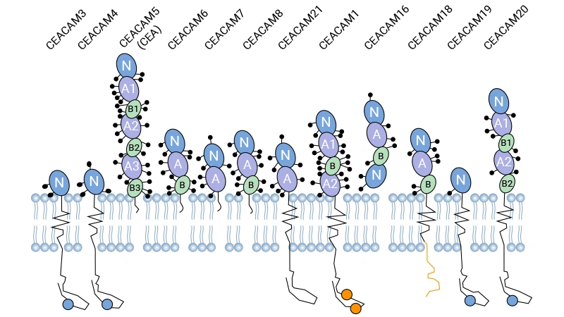
Figure. Schematic structures of major CEACAM family members [1]
Among them, CEACAM1, CEACAM3, CEACAM5, CEACAM6, CEACAM7, and CEACAM8 are the most extensively studied core members. Most CEACAM proteins contain an N-terminal IgV-like domain together with varying numbers of IgC2-like domains, enabling homophilic or heterophilic cell adhesion. In terms of membrane association, CEACAM1 and CEACAM3 are transmembrane proteins, whereas CEACAM5, CEACAM6, CEACAM7, and CEACAM8 are primarily anchored to the cell membrane through glycosylphosphatidylinositol (GPI).
Different CEACAM members also differ in their intracellular domains and membrane-anchoring patterns. For example, CEACAM1 contains a typical immunoreceptor tyrosine-based inhibitory motif (ITIM), whereas CEACAM3 exhibits ITAM-like signaling features. In contrast, several GPI-anchored members lack obvious intracellular signaling domains.
CEACAM family members exhibit substantial functional diversity, with major roles in cell adhesion, signal regulation, immune modulation, and host–pathogen interactions. Many CEACAM proteins participate in cell aggregation, maintenance of cellular polarity, and cell migration through homophilic or heterophilic interactions, while some members additionally regulate downstream immune- or inflammation-related signaling pathways.
Beyond their cellular biological functions, CEACAM family members are also widely involved in host–pathogen interactions. Numerous bacteria and viruses have been reported to exploit CEACAM molecules to enhance host adhesion or modulate immune responses, which has made the CEACAM family an important focus in infection biology research.
CEACAM family members display distinct tissue distribution patterns. CEACAM5, CEACAM6, and CEACAM7 are mainly expressed in epithelial tissues of the gastrointestinal and respiratory tracts. Among them, CEACAM5 is frequently upregulated in a variety of epithelial-derived tumors, whereas CEACAM7 is often downregulated or lost in colorectal cancer.
CEACAM1, CEACAM3, CEACAM6, and CEACAM8 can be expressed in neutrophils and certain immune cell populations. CEACAM3 and CEACAM8 show more restricted expression within granulocyte-related systems, whereas CEACAM1 displays broader tissue distribution, including epithelial cells, endothelial cells, and multiple immune cell types.
CEACAM6 exhibits a relatively unique expression profile. In addition to its high expression in multiple epithelial-derived tumors, it can also be detected in neutrophils, thereby linking both tumor biology and inflammation-related research contexts.
Based on differences in tissue distribution and functional characteristics, research on the CEACAM family has gradually developed into several major directions.
Among these members, CEACAM5, CEACAM6, and CEACAM7 are mainly studied in the context of epithelial biology and cancer; CEACAM3 and CEACAM8 are more closely associated with granulocyte function and host defense; while CEACAM1, which contains inhibitory signaling features, is broadly involved in immune regulation and host–pathogen interactions.
In addition, because some pathogens can bind multiple CEACAM members simultaneously, the CEACAM family has increasingly become an important link connecting cancer biology, immunology, and infection research.
The complexity of tumor immune evasion mechanisms lies in their multi-layered inhibitory network. Therefore, research decisions must start from a macro perspective to screen for Siglec targets with strategic complementarity. Current evidence indicates that the sialic acid-Siglec axis constructs a well-defined immunosuppressive system through the specific expression of different molecules on particular immune cell subsets. Based on this, research can focus on the following three core directions:
CEACAM5 (classical carcinoembryonic antigen, CEA) is one of the most extensively studied and clinically utilized CEACAM family members. In epithelial-derived tumors such as colorectal cancer, gastric cancer, non-small cell lung cancer, and pancreatic cancer, CEACAM5 expression is frequently elevated and has been widely used for auxiliary diagnosis, therapeutic monitoring, and recurrence assessment [2–4]. In normal adult tissues, CEACAM5 expression is generally restricted to polarized epithelial surfaces, whereas tumor tissues often exhibit both increased expression and loss of polarity. Beyond its role as a serum biomarker, membrane-associated CEACAM5 has also been implicated in abnormal tumor cell adhesion and local tumor dissemination.
Compared with CEACAM5, CEACAM6 research is more strongly focused on tumor progression-related functions. Multiple studies have demonstrated that elevated CEACAM6 expression is associated with malignant phenotypes, including anoikis resistance, enhanced invasion and migration, chemoresistance, and epithelial–mesenchymal transition (EMT). High CEACAM6 expression is frequently associated with poor prognosis in pancreatic cancer, breast cancer, colorectal cancer, and lung cancer [5].
In recent years, CEACAM6 has also attracted increasing attention in translational medicine. Several studies have begun exploring CEACAM6-based antibody-drug conjugates (ADCs) and targeted delivery strategies aimed at improving selective drug delivery to tumors with high CEACAM6 expression [14]. This trend suggests that CEACAM6 is gradually evolving from a "tumor-associated molecule" into a therapeutic target with broader translational potential.
Compared with CEACAM5 and CEACAM6, CEACAM7 has been studied less extensively, although its unique expression pattern remains biologically informative. CEACAM7 is relatively stably expressed in normal colonic epithelium but is frequently downregulated or lost in colorectal cancer tissues. As a result, it is often used to evaluate alterations in epithelial differentiation status and tumor-associated expression abnormalities. Unlike CEACAM5 and CEACAM6, which are generally characterized by increased expression, changes in CEACAM7 more commonly reflect disruption of epithelial homeostasis and differentiation.
More recently, several studies have investigated aberrant CEACAM7 expression in pancreatic ductal adenocarcinoma (PDAC) and explored its potential as an immunotherapeutic target. For example, CEACAM7 expression has been detected on the surface of subsets of PDAC cells and has therefore been considered in exploratory CAR-T targeting strategies [6]. However, the functional mechanisms and therapeutic relevance of CEACAM7 remain insufficiently characterized. Accordingly, CEACAM7 is currently better regarded as a supportive member for understanding alterations in epithelial CEACAM expression profiles rather than a central functional molecule equivalent to CEACAM6.
In epithelial tumor research, simultaneous evaluation of CEACAM5/6 upregulation and CEACAM7 downregulation may provide a more comprehensive understanding of CEACAM-associated expression changes related to tumor progression and differentiation status.
CEACAM3 is one of the most host defense-oriented members of the CEACAM family and is primarily expressed on neutrophils in humans and certain primates. Its intracellular domain contains an ITAM-like motif capable of recruiting Syk kinase following receptor activation, thereby inducing phagocytosis, oxidative burst, and inflammatory signaling activation [7,8].
Unlike classical Fc receptors that depend on antibody- or complement-mediated opsonization, CEACAM3 can directly recognize pathogens expressing CEACAM-binding adhesins, including Neisseria gonorrhoeae, Neisseria meningitidis, and certain Moraxella species, thereby promoting rapid neutrophil-mediated phagocytosis. Consequently, CEACAM3 is widely considered an important host defense mechanism against CEACAM-binding pathogens in human neutrophils.
CEACAM8 (CGM6) is one of the highly expressed CEACAM members in neutrophils and is closely associated with granulocyte maturation and activation status. Unlike many transmembrane CEACAM proteins, CEACAM8 is a GPI-anchored protein that can be released into the extracellular environment during neutrophil activation and degranulation. As a result, it is frequently used as an indicator of granulocyte activation and inflammatory status [9].
Recent studies have reported elevated soluble CEACAM8 levels in chronic airway inflammation, sepsis, and certain autoimmune diseases, suggesting its potential utility as an auxiliary biomarker reflecting neutrophil activation and inflammatory responses. Compared with CEACAM3, which functions primarily as a host defense receptor, CEACAM8 is more closely associated with inflammatory state evaluation and soluble readout-related studies.
In addition to the classical granulocyte-associated members, some studies have also focused on the role of CEACAM6 in inflammation and host–pathogen interactions. Several studies have demonstrated that Opa protein-expressing Neisseria species can interact with both CEACAM3 and CEACAM6. In this context, CEACAM3 appears to be more directly associated with phagocytic activation, whereas CEACAM6 may contribute to cell adhesion, receptor clustering, and inflammation-related responses [10,11].
Because CEACAM6 is expressed in both epithelial cells and neutrophils, its biological significance extends beyond pathogen adhesion to include cellular interactions within inflammatory microenvironments. In addition, some studies have suggested that soluble CEACAM6 may participate in inflammation-associated signaling regulation, although its precise mechanisms remain incompletely understood. Accordingly, in granulocyte-related research, CEACAM6 is better regarded as a supportive molecule linking inflammatory responses and host–pathogen interactions rather than as a classical phagocytic receptor comparable to CEACAM3.
Multiple pathogens, including pathogenic Escherichia coli, Salmonella, Haemophilus influenzae, and Helicobacter pylori, have been reported to interact with different CEACAM family members. Among them, the Opa proteins of Neisseria gonorrhoeae can bind CEACAM1, CEACAM3, CEACAM5, and CEACAM6, thereby enhancing adhesion to epithelial cells while also modulating host immune responses [11].
Importantly, different CEACAM members are expressed in distinct cell types and possess different signaling properties. As a result, pathogen interactions with different CEACAM proteins may lead to distinct biological outcomes.
CEACAM1 is one of the most important immunoregulatory members of the CEACAM family. Its intracellular domain contains an immunoreceptor tyrosine-based inhibitory motif (ITIM). Upon receptor ligation or crosslinking, the ITIM becomes phosphorylated and recruits phosphatases such as SHP-1 and SHP-2, thereby suppressing downstream activation signaling and negatively regulating T cells, NK cells, and certain myeloid cell populations.
Because CEACAM1 possesses characteristics of both an adhesion molecule and an inhibitory receptor, it has received considerable attention in tumor immunology, inflammatory regulation, and host–pathogen interaction studies. Within the tumor microenvironment, CEACAM1-mediated homophilic or heterophilic interactions have been associated with immune inhibitory signaling and T-cell exhaustion phenotypes [12]. Some studies further suggest that sustained CEACAM1 expression may contribute to tumor immune evasion by reducing local immune-mediated cytotoxicity [13].
At the same time, several pathogens can exploit CEACAM1 as a binding receptor to influence host cell signaling and inflammatory responses. Consequently, CEACAM1 is widely regarded as a key bridge connecting tumor biology, mucosal immunity, and host–pathogen interactions.
Differences in expression patterns, tissue distribution, and functional specialization among CEACAM family members strongly influence experimental strategy selection. In general, current CEACAM-related studies can be categorized into three major areas: expression and association analysis, soluble level quantification, and functional or mechanistic investigation. Because different research questions require distinct experimental readouts and reagents, defining the central scientific objective is often more important than selecting a specific technical platform.
When the primary objective involves tissue expression, cellular localization, or correlations with clinicopathological parameters, antibody-based detection approaches are typically central. Immunohistochemistry, Western blotting, and flow cytometry remain among the most widely used methods. Immunohistochemistry is particularly useful for evaluating spatial distribution and expression polarity in tissues, whereas flow cytometry is better suited for analyzing membrane-associated CEACAM expression on immune cells or tumor cells.
For epithelial-associated members such as CEACAM5 and CEACAM6, expression studies generally focus on abnormal upregulation and altered polarity, often combined with analyses of tumor stage, invasiveness, or treatment response. In contrast, studies involving CEACAM3 and CEACAM8 are more frequently associated with granulocyte activation status, inflammatory conditions, or pathogen stimulation.
Because extracellular domains among CEACAM family members exhibit substantial homology, antibody cross-reactivity remains an important experimental consideration. Therefore, studies involving comparisons among multiple CEACAM members typically require carefully validated antibody systems. In addition, glycosylation-related molecular weight shifts and epitope accessibility differences should also be considered when interpreting experimental data.
In addition to membrane-associated expression, several CEACAM family members can also be released into the extracellular environment, making soluble level analysis an important area of investigation. Compared with tissue expression studies, these analyses are more closely associated with dynamic changes, disease activity, and inflammation-related readouts.
ELISA remains one of the most commonly used strategies for quantitative analysis in serum, plasma, and cell culture supernatants. CEACAM5 (CEA) has long been utilized as a clinical tumor biomarker, whereas CEACAM6 and CEACAM8 are more frequently investigated in the context of inflammation, granulocyte activation, and host response studies. In recent years, multiplex detection systems and electrochemiluminescence-based platforms have also increasingly been applied to CEACAM-related studies to support complex sample analysis and multi-marker profiling.
Importantly, the biological significance of soluble CEACAMs is not always equivalent to tissue expression. For example, CEACAM6 may exist in both membrane-bound and soluble forms in certain tumors. Therefore, integrating tissue expression data with soluble level measurements may provide a more comprehensive understanding of disease-associated dynamic changes.
When research objectives extend beyond expression analysis toward cellular behavior and signaling mechanisms, the primary focus shifts from "expression presence" to "functional involvement." Such studies typically investigate the effects of CEACAM members on proliferation, migration, apoptosis, phagocytosis, inflammatory signaling, and intercellular interactions.
In mechanistic studies, recombinant proteins are commonly used to model receptor binding, cell adhesion, or ligand competition processes, whereas functional antibodies may be employed to block or activate specific CEACAM-mediated signaling pathways. For example, CEACAM6 is frequently investigated in relation to invasion, anoikis resistance, and therapeutic resistance in tumor biology, while CEACAM3 is more commonly studied in pathogen recognition, phagocytic activation, and inflammatory output in granulocyte research.
Furthermore, because CEACAM1 and CEACAM3 contain ITIM- and ITAM-like-associated signaling features, respectively, downstream phosphorylation events and signaling complex formation are also major areas of mechanistic investigation. However, significant differences may exist among experimental model systems in terms of expression profiles and signaling integrity; therefore, findings should always be interpreted within the context of the specific cellular background used.
To support CEACAM family-related research, CUSABIO provides recombinant proteins, antibodies, and ELISA kits suitable for expression analysis, functional studies, and soluble level quantification across multiple research scenarios, including tumor biology, granulocyte function, and host–pathogen interaction studies.
| Target | Code | Product Name | Source |
|---|---|---|---|
| CEACAM1 | CSB-EP005157HU | Recombinant Human Carcinoembryonic antigen-related cell adhesion molecule 1 (CEACAM1), partial | E.coli |
| CEACAM1 | CSB-EP005157HUe1 | Recombinant Human Carcinoembryonic antigen-related cell adhesion molecule 1 (CEACAM1), partial | E.coli |
| CEACAM1 | CSB-MP005157HU-B | Recombinant Human Carcinoembryonic antigen-related cell adhesion molecule 1 (CEACAM1), partial, Biotinylated (Active) | Mammalian cell |
| CEACAM1 | CSB-EP005157HUd7 | Recombinant Human Carcinoembryonic antigen-related cell adhesion molecule 1 (CEACAM1), partial | E.coli |
| CEACAM1 | CSB-YP005157HU | Recombinant Human Cell adhesion molecule CEACAM1 (CEACAM1), partial (Active) | Yeast |
| CEACAM3 | CSB-EP005163HU | Recombinant Human Carcinoembryonic antigen-related cell adhesion molecule 3 (CEACAM3), partial | E.coli |
| CEACAM4 | CSB-EP005164HU | Recombinant Human Cell adhesion molecule CEACAM4 (CEACAM4), partial | E.coli |
| CEACAM4 | CSB-YP005164HU | Recombinant Human Cell adhesion molecule CEACAM4 (CEACAM4), partial | Yeast |
| CEACAM4 | CSB-MP005164HU | Recombinant Human Cell adhesion molecule CEACAM4 (CEACAM4), partial | Mammalian cell |
| CEACAM5 | CSB-YP005165HU | Recombinant Human Carcinoembryonic antigen-related cell adhesion molecule 5 (CEACAM5) (E398K) | Yeast |
| CEACAM5 | CSB-MP005165HU | Recombinant Human Carcinoembryonic antigen-related cell adhesion molecule 5 (CEACAM5) (E398K) (Active) | Mammalian cell |
| Ceacam5 | CSB-MP005165MO(A4) | Recombinant Mouse Carcinoembryonic antigen-related cell adhesion molecule 5 (Ceacam5) | Mammalian cell |
| CEACAM5 | CSB-MP5152MOW | Recombinant Macaca mulatta Carcinoembryonic antigen-related cell adhesion molecule 5 (CEACAM5), partial (Active) | Mammalian cell |
| CEACAM5 | CSB-MP005165HUd9 | Recombinant Human Cell adhesion molecule CEACAM5 (CEACAM5), partial | Mammalian cell |
| CEACAM5 | CSB-MP005165HU(M)-B | Recombinant Human Cell adhesion molecule CEACAM5 (CEACAM5) (E398K), Biotinylated | Mammalian cell |
| CEACAM6 | CSB-EP005166HU | Recombinant Human Carcinoembryonic antigen-related cell adhesion molecule 6 (CEACAM6) | E.coli |
| CEACAM6 | CSB-MP005166HU | Recombinant Human Carcinoembryonic antigen-related cell adhesion molecule 6 (CEACAM6) (Active) | Mammalian cell |
| CEACAM7 | CSB-EP005167HU | Recombinant Human Carcinoembryonic antigen-related cell adhesion molecule 7 (CEACAM7) | E.coli |
| CEACAM8 | CSB-EP005168HU | Recombinant Human Carcinoembryonic antigen-related cell adhesion molecule 8 (CEACAM8) | E.coli |
| CEACAM8 | CSB-MP005168HU | Recombinant Human Carcinoembryonic antigen-related cell adhesion molecule 8 (CEACAM8) (Active) | Mammalian cell |
| Target | Code | Product Name | Tested Applications |
|---|---|---|---|
| CEACAM1 | CSB-RA147192A0HU | CEACAM1 Recombinant Monoclonal Antibody | ELISA, IHC |
| CEACAM1 | CSB-PA005134 | CEACAM1 Antibody | WB, ELISA |
| CEACAM1 | CSB-PA088258 | CEACAM1 Antibody | ELISA, WB, IHC |
| CEACAM1 | CSB-PA566314 | CEACAM1 Antibody | ELISA, WB |
| CEACAM1 | CSB-PA815431 | CEACAM1 Antibody | ELISA, WB, IHC |
| CEACAM1 | CSB-PA939516 | CEACAM1 Antibody | ELISA, WB, IHC |
| CEACAM1 | CSB-PA10749A0Rb | CEACAM1 Antibody | ELISA, WB, IF |
| CEACAM1 | CSB-PA10749B0Rb | CEACAM1 Antibody, HRP conjugated | ELISA |
| CEACAM1 | CSB-PA10749C0Rb | CEACAM1 Antibody, FITC conjugated | N/A |
| CEACAM1 | CSB-PA10749D0Rb | CEACAM1 Antibody, Biotin conjugated | ELISA |
| CEACAM3 | CSB-PA260295 | CEACAM3 Antibody | ELISA, WB, IHC |
| CEACAM3 | CSB-PA055165 | CEACAM3 Antibody | ELISA, IHC |
| CEACAM3 | CSB-PA795702 | CEACAM3 Antibody | ELISA, IHC |
| CEACAM3 | CSB-PA965136 | CEACAM3 Antibody | ELISA, IHC |
| CEACAM3 | CSB-PA005163ESR2HU | CEACAM3 Antibody | ELISA, IHC |
| CEACAM3/CEACAM6 | CSB-PA006209 | CEACAM3/CEACAM6 Antibody | WB, ELISA |
| CEACAM4 | CSB-PA005164LA01HU | CEACAM4 Antibody | ELISA |
| CEACAM4 | CSB-PA005164LB01HU | CEACAM4 Antibody, HRP conjugated | ELISA |
| CEACAM4 | CSB-PA005164LC01HU | CEACAM4 Antibody, FITC conjugated | N/A |
| CEACAM4 | CSB-PA005164LD01HU | CEACAM4 Antibody, Biotin conjugated | ELISA |
| Target | Code | Product Name | Detection Range | Sensitivity |
|---|---|---|---|---|
| CEACAM1 | CSB-EL005157HU | Human Carcinoembryonic antigen-related cell adhesion molecule 1(CEACAM1) ELISA kit | 0.78 ng/mL-50 ng/mL | 0.195 ng/mL |
| CEACAM5 | CSB-E04767h | Human carcinoembryonic antigen,CEA ELISA Kit | 5 ng/ml-80 ng/ml | 2 ng/mL |
| CEACAM5 | CSB-E13925m | Mouse carcinoembryonic antign,CEA ELISA Kit | 62.5 pg/mL-4000 pg/mL | 15.6 pg/mL |
| CEACAM6 | CSB-EL005166HU | Human Carcinoembryonic antigen-related cell adhesion molecule 6(CEACAM6) ELISA kit | 0.83 ng/mL-20 ng/mL | 0.33 ng/mL |
| CEACAM8 | CSB-EL005168HU | Human Carcinoembryonic antigen-related cell adhesion molecule 8(CEACAM8) ELISA kit | 0.625 ng/mL-40 ng/mL | 0.156 ng/mL |
[1] Johnson B,Mahadevan D. Emerging Role and Targeting of Carcinoembryonic Antigen-related Cell Adhesion Molecule 6 (CEACAM6) in Human Malignancies. Clin Cancer Drugs. 2015;2 (2):100-111.
[2] Kopetz S,Boni V,Kato K, et al. Precemtabart tocentecan, an anti-CEACAM5 antibody-drug conjugate, in metastatic colorectal cancer: a phase 1 trial. Nat Med. 2025;31 (10):3504-3513.
[3] Zhou J,Fan X,Chen N, et al. Identification of CEACAM5 as a Biomarker for Prewarning and Prognosis in Gastric Cancer. J Histochem Cytochem. 2015;63 (12):922-30.
[4] Shi H,Tsang Y,Yang Y. Identification of CEACAM5 as a stemness-related inhibitory immune checkpoint in pancreatic cancer. BMC Cancer. 2022;22 (1):1291.
[5] Wu G,Wang D,Xiong F, et al. The emerging roles of CEACAM6 in human cancer (Review). Int J Oncol. 2024;64 (3):.
[6] Raj D,Nikolaidi M,Garces I, et al. CEACAM7 Is an Effective Target for CAR T-cell Therapy of Pancreatic Ductal Adenocarcinoma. Clin Cancer Res. 2021;27 (5):1538-1552.
[7] Bonsignore P,Kuiper JWP,Adrian J, et al. CEACAM3-A Prim(at)e Invention for Opsonin-Independent Phagocytosis of Bacteria. Front Immunol. 2019;10:3160.
[8] Kuiper JWP,Gregg HL,Schüber M, et al. Controling the cytoskeleton during CEACAM3-mediated phagocytosis. Eur J Cell Biol. 2024;103 (1):151384.
[9] Ribon M,Mussard J,Semerano L, et al. Extracellular Chromatin Triggers Release of Soluble CEACAM8 Upon Activation of Neutrophils. Front Immunol. 2019;10:1346.
[10] Strickland LA,Ross J,Williams S, et al. Preclinical evaluation of carcinoembryonic cell adhesion molecule (CEACAM) 6 as potential therapy target for pancreatic adenocarcinoma. J Pathol. 2009;218 (3):380-90.
[11] Islam EA,Anipindi VC,Francis I, et al. Specific Binding to Differentially Expressed Human Carcinoembryonic Antigen-Related Cell Adhesion Molecules Determines the Outcome of Neisseria gonorrhoeae Infections along the Female Reproductive Tract. Infect Immun. 2018;86 (8).
[12] Kim WM,Huang YH,Gandhi A, et al. CEACAM1 structure and function in immunity and its therapeutic implications. Semin Immunol. 2019;42:101296.
[13] Dankner M,Gray-Owen SD,Huang YH, et al. CEACAM1 as a multi-purpose target for cancer immunotherapy. Oncoimmunology. 2017;6 (7):e1328336.
[14] Tsai YT,Hu PJ,Liu CY, et al. A heavy-chain antibody-drug conjugate targeting glycosylated CEACAM6 inhibits brain metastatic tumor growth. Cancer Lett. 2026;648:218463.