

Call us
301-363-4651 (Available 9 a.m. to 5 p.m. CST from Monday to Friday)
Cytokines are soluble signaling proteins that orchestrate intercellular communication across innate and adaptive immunity, inflammation, tissue homeostasis, and disease pathogenesis. Accurate, sensitive, and high-throughput detection of cytokines is foundational to basic immunological research, biomarker discovery, and the development of novel therapeutics for cancer, autoimmune disorders, infectious diseases, and inflammatory conditions.
This article provides a comprehensive overview of established and emerging technologies for cytokine detection, from gold-standard immunoassays to cutting-edge single-molecule, single-cell, and spatial profiling platforms. We detail the working principles, advantages, and limitations of each methodology and also provide practical guidance on pre-analytical and analytical best practices to ensure robust and reproducible cytokine measurements. Finally, we highlight translational applications of these technologies and discuss future directions in the field, equipping researchers with the knowledge to select the optimal detection strategy for their experimental and clinical research goals.
Table of Contents
2. Why Do We Detect Cytokines?
3. Technologies for Cytokine Detection
4. Emerging Biosensing Technologies for Cytokine Detection
5. Critical Considerations in Cytokine Measurement
6. Translational and Clinical Applications of Cytokine Detection
Cytokines are a diverse superfamily of soluble proteins, including interleukins (ILs), interferons (IFNs), tumor necrosis factors (TNFs), colony-stimulating factors (CSFs), chemokines, and growth factors, that mediate cell-cell communication in nearly all biological processes. Produced transiently and in a cell-specific manner in response to environmental stimuli, cytokines act via autocrine, paracrine, and endocrine signaling to regulate immune cell activation, differentiation, proliferation, and apoptosis.
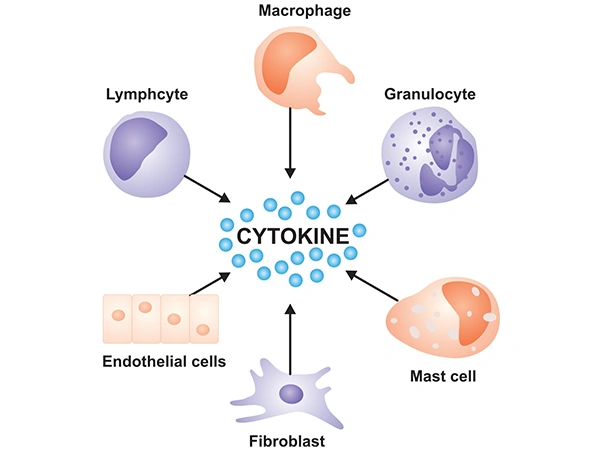
Figure 1. Various cells that produce cytokines
Cytokines exert biological effects by binding to corresponding cytokine receptors on the cell surface. The combination of cytokines and their receptors initiates complex intracellular molecular interactions, ultimately causing changes in cellular gene transcription.
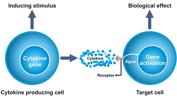
Figure 2. The process by which cytokines work
Cytokines are soluble, low-molecular-weight signaling proteins that orchestrate intercellular communication within the immune system, regulating critical processes including inflammation, immune cell maturation, pathogen defense, and tissue homeostasis.
Dysregulation of cytokine signaling is a hallmark of a broad spectrum of human diseases, including rheumatoid arthritis, multiple sclerosis, cancer, sepsis, and neurodegenerative disorders. Under physiological conditions, circulating cytokines are typically present at picomolar to low picogram-per-milliliter concentrations, but their levels can increase up to 1,000-fold during immune activation or pathological dysregulation. As such, the ability to precisely quantify cytokine expression and secretion is indispensable for decoding immune regulatory networks, identifying disease biomarkers, and evaluating the efficacy of immunotherapies and vaccines.
These foundational methodologies remain the backbone of cytokine research, with well-validated protocols, broad accessibility, and well-defined performance characteristics for routine laboratory use. The best method depends on whether the goal is absolute quantification, multiplex profiling, single-cell resolution, or near-real-time monitoring.
ELISA remains a classic method for cytokine measurement because it is familiar, relatively straightforward, and well-established. It is often used for single-analyte quantification in serum, plasma, or culture supernatants.
The sandwich ELISA is the most common method for cytokine quantification. It uses matched antibody pairs that bind to different epitopes of the target cytokine. The cytokine in the sample is captured by an immobilized antibody, detected by a second antibody, and then measured through an enzyme-conjugated antibody and colorimetric or chemiluminescent signal readout. The signal intensity, quantified against a standard curve, is proportional to the cytokine concentration.
.webp)
Figure 3. Sandwich ELISA (Indiect)
- Chemiluminescent ELISA: Replaces colorimetric substrates with chemiluminescent reagents, improving LOD by 10–100 fold compared to colorimetric ELISA [2].
- Electrochemical ELISA: Uses electrochemical signal transduction, enabling miniaturization and point-of-care (POC) applications [6].
Given that cytokines act in complex, interconnected networks, multiplex methods that simultaneously quantify dozens of cytokines in a single small-volume sample have become foundational for translational and clinical research.
These assays use spectrally encoded microspheres (beads) conjugated to cytokine-specific capture antibodies. Each bead population has a unique fluorescent signature, enabling target differentiation in a single reaction. After incubation with the sample and fluorophore-labeled detection antibodies, bead fluorescence is quantified via flow cytometry or dedicated analyzers [1][2].
- Luminex xMAP Technology: Supports up to 100 analytes per well, with a sample volume requirement of 25–50 μL, and a 3–4 order of magnitude linear dynamic range [1][7].
- Cytometric Bead Array (CBA): Compatible with standard laboratory flow cytometers, lowering the barrier to entry for multiplex cytokine analysis [2][7].
- One-Step Flow Cytometry-Based Multiplex Assays: Lyophilized, room-temperature-stable reagent mixes that reduce assay time and cold-chain requirements, validated on clinical serum samples from patients with infectious diseases [3].
Microarrays use spatially patterned capture antibodies printed on glass or membrane slides, enabling simultaneous detection of up to hundreds of cytokines in a single sample. Signal detection is typically via fluorescence or chemiluminescence, with dedicated scanners for quantification [1][2].
Bulk cytokine detection methods cannot resolve cellular heterogeneity, which is critical for identifying rare cytokine-producing cell populations (e.g., antigen-specific T cells) in immune monitoring. The following methods enable single-cell resolution cytokine analysis [1][2].
ELISPOT is the gold-standard method for quantifying the frequency of cytokine-secreting cells at the single-cell level. The assay uses capture antibodies coated on a microplate well to trap cytokines secreted by individual live cells cultured on the plate. Captured cytokines are then detected by enzyme-conjugated secondary antibodies, forming visible spots that correspond to individual cytokine-producing cells [1][2].
ICS combines flow cytometry with immunofluorescent staining to detect cytokines inside fixed and permeabilized cells, enabling simultaneous measurement of cytokine production and cell surface immunophenotype markers (e.g., CD4, CD8) at the single-cell level [1][2].
Conventional immunoassays lack the sensitivity to detect low, homeostatic cytokine concentrations in healthy individuals, which are critical for establishing baseline levels and early disease detection. Ultrasensitive technologies achieve LODs in the sub-pg/mL to fg/mL range by counting single cytokine molecules digitally [2][5].
Simoa is a digital ELISA platform that uses antibody-conjugated magnetic microbeads to capture cytokines, followed by immunocomplex formation with enzyme-labeled detection antibodies. Individual beads are isolated in femtoliter-scale microwells, where fluorogenic substrate hydrolysis generates a fluorescent signal in wells containing a single target cytokine molecule [2][5].
These methods indirectly measure cytokine production by quantifying cytokine mRNA transcripts, providing insights into cellular cytokine gene expression rather than secreted protein levels [1][2].
qRT-PCR is the standard method for quantifying cytokine mRNA expression in cell or tissue samples, using reverse transcription to convert cytokine mRNA to cDNA, followed by real-time PCR amplification and fluorescence-based quantification [1][2].
scRNA-seq enables transcriptome-wide profiling of cytokine gene expression at the single-cell level, resolving the cellular heterogeneity of cytokine production in complex immune cell populations [2].
Mass spectrometry (MS) methods enable antibody-independent, label-free detection and quantification of cytokines, with the ability to simultaneously measure cytokine isoforms, post-translational modifications, and degradation products [2][4].
Key Approaches:
Conventional cytokine detection methods are largely limited to centralized laboratory settings, requiring bulky instrumentation, specialized expertise, long turnaround times, and large sample volumes [1][2][6]. Emerging biosensing technologies integrate nanomaterial engineering, microfluidics, and molecular tools to transduce cytokine binding events into quantifiable signals, enabling rapid, miniaturized, POC, and continuous cytokine detection with ultra-high sensitivity and minimal sample consumption [2][6][10].
Electrochemical biosensors are the most clinically advanced emerging cytokine detection platform, converting cytokine-antibody/aptamer binding into measurable electrical signals (amperometric, voltammetric, impedimetric) via a sandwich immunoassay format on a functionalized electrode surface [10][11].
Optical biosensors detect cytokines by measuring light property changes induced by target binding to immobilized capture probes, with surface plasmon resonance (SPR), localized SPR (LSPR), and surface-enhanced Raman scattering (SERS) as the dominant modalities [7][12][13].
FET biosensors are label-free electrical platforms that transduce cytokine binding directly into current changes via a semiconductor channel functionalized with capture probes [17].
Microfluidic "lab-on-a-chip" platforms integrate cytokine detection with microscale fluidic control, automating all assay steps (sample processing, incubation, detection) on a single chip [14].
These platforms address the need for low-cost, accessible cytokine monitoring outside centralized labs, with two core formats:
CRISPR-enhanced platforms leverage the collateral cleavage activity of Cas12a/Cas13a systems to amplify cytokine binding signals, combining antibody-based capture with CRISPR-mediated signal amplification [18][24].
A cytokine assay is only as reliable as the design and handling behind it. Many apparent "biological" differences are actually caused by sampling, storage, or platform-related variation.
Sensitivity determines whether a method can detect low concentrations, while specificity determines whether it measures the intended cytokine rather than a related molecule. Antibody quality, assay format, and cross-reactivity are central to both [27,28].
This is why platform validation matters. Even strong technologies can give misleading results if capture and detection reagents are poorly matched or if the cytokine concentration is near the assay’s lower limit [28,29].
Cytokine assays often vary across laboratories, kits, and analysis pipelines. Without standardized procedures and reference controls, comparison between studies can become difficult [30,31].
Reproducibility improves when the assay conditions are tightly defined, including incubation time, temperature, vessel type, and readout method. One ex vivo stimulation study found that temperature control and stimulation timing were among the most important sources of variability [30].
Pre-analytical factors account for up to 70% of the variability in cytokine measurement data, making them the single most important determinant of data reproducibility. Pre-analytical factors include sample type selection, sample collection, transport, centrifugation, storage, freeze-thaw cycles, and the use of different collection tubes. These steps can change cytokine levels before analysis even begins [31,32].
Many studies agree that pre-analytical control is essential. In practical terms, this means using consistent workflows, minimizing delays, and documenting every step from blood draw to assay plate [31,32].
Cytokine data often need normalization, especially in multiplex or longitudinal studies. Batch effects, matrix effects, and biological variability can all distort the final picture if they are not addressed [33,34].
Interpretation should also be cautious. A cytokine increase may reflect inflammation, but it may also reflect cell composition, sample timing, or treatment effects. Good analysis usually combines cytokine data with clinical variables, cell counts, or functional assays [35,36].
Cytokine measurement is not limited to basic research. It is increasingly used to support diagnosis, monitor disease course, and guide therapy in settings where immune activity matters.
Cytokines can serve as biomarkers for diagnosis, prognosis, and patient stratification. They are being explored in cancer, autoimmune disease, infection, and inflammatory syndromes because they reflect immune activity in a direct way [36,37].
That said, cytokines are rarely specific on their own. Their value often comes from being part of a panel or signature rather than a single, standalone marker [36,37].
In drug development, cytokine monitoring can help evaluate pharmacodynamic response and immune activation. This is especially important for therapies that alter immune signaling, such as checkpoint inhibitors or CAR-T cells [30,34].
Studies using cytokine profiling during CAR-T treatment have shown that multiplex and digital assays can track evolving immune states over time. This makes cytokines useful both as mechanistic readouts and as safety-related monitoring tools [34,38].
Cytokine testing is widely used in severe infection, sepsis, and viral disease research. In these settings, high levels of cytokines may reflect hyperinflammation or cytokine storm, which can help identify high-risk patients [33,36].
COVID-19 studies have shown that panels of circulating cytokines can help distinguish patient subgroups and track disease severity. Similar principles apply to other inflammatory diseases, where timing and trajectory matter as much as the absolute value [33,39].
Cytokine profiles can support more individualized treatment decisions by showing which inflammatory pathways are active in a given patient. This is most useful when the profile is interpreted together with other biomarkers and clinical data [35,40].
In practice, personalized medicine depends on longitudinal measurement, careful validation, and clinically meaningful thresholds. Cytokines are promising here because they can reflect dynamic immune changes in real time, but they still need to be used with discipline [35,36].
Despite dramatic advances in cytokine detection technologies, significant challenges remain, driving ongoing innovation in the field. The most important challenge is not just making assays more sensitive but also more robust, comparable, and clinically useful.
Current limitations include cross-reactivity, cost, restricted dynamic range, and differences between assay platforms. These issues can complicate both research interpretation and clinical deployment [28,29].
Even advanced multiplex or digital systems must still deal with matrix effects and calibration challenges. As assays become more complex, careful validation becomes more—not less—important [34].
Cytokine data become more informative when combined with transcriptomics, proteomics, metabolomics, and cell phenotyping. This systems-level view can help explain why a cytokine signal appears, which cells contribute to it, and what it means biologically [41,42].
Multi-omics integration is especially promising for biomarker discovery and disease stratification. It can also reduce overreliance on any single cytokine as a stand-alone marker [35,42].
Researchers are increasingly interested in rapid and near-real-time cytokine monitoring. Microfluidic and biosensor systems are moving in this direction by shortening assay time and reducing the amount of sample needed [38].
The long-term goal is not just faster testing, but continuous or minimally invasive readouts that can track immune status as it changes. That would be especially valuable in critical care, immunotherapy, and infectious disease.
For cytokine assays to move deeper into clinical use, they need common standards, transparent validation, and reproducible workflows. Without this, results may remain difficult to compare across platforms and institutions [31,40].
Standardization is particularly important for biomarker-driven decisions, where small analytic differences may affect treatment interpretation. The more clinically relevant the use case, the higher the bar for assay consistency [35,40].
Cytokine detection has moved from simple single-analyte measurement to multiplex, single-cell, and digital profiling. Each technology has distinct advantages and limitations, and the optimal detection strategy depends on the research question, sample type, required sensitivity, and multiplexing capacity.
For researchers, the best approach is to match the method to the biological question. For clinicians and translational teams, the next step is to turn cytokine data into reliable, standardized, and actionable information.
As technology continues to advance, cytokine detection will become increasingly sensitive, high-throughput, and physiologically relevant, enabling unprecedented insights into cytokine signaling networks and the development of novel cytokine-targeted therapeutics for a broad spectrum of human diseases.
References
[1] Stenken, J. A., & Poschenrieder, A. J. (2014). Bioanalytical Chemistry of Cytokines-A Review [J]. Analytica Chimica Acta, 853, 95.
[2] Liu, C., Chu, D., George, J., Young, H. A., & Liu, G. (2021). Cytokines: From Clinical Significance to Quantification [J]. Advanced Science, 8(15), 2004433.
[3] Quan, Q., Ju, X., et al. (2025). Simplified flow cytometry-based assay for rapid multi-cytokine profiling and machine-learning-assisted diagnosis of inflammatory diseases [J]. Frontiers in Pharmacology, 16, 1594141.
[4] Skalnikova, H. K., Cizkova, J., Cervenka, J., & Vodicka, P. (2017). Advances in Proteomic Techniques for Cytokine Analysis: Focus on Melanoma Research [J]. International Journal of Molecular Sciences, 18(12), 2697.
[5] McKinski, K., Tang, H., Wang, K., Birchler, M., & Wright, M. (2024). Comparison of highly sensitive, multiplex immunoassay platforms for streamlined clinical cytokine quantification [J]. Bioanalysis, 17(1), 17.
[6] Liu, G., Qi, M., Hutchinson, M. R., Yang, G., & Goldys, E. M. (2016). Recent advances in cytokine detection by immunosensing [J]. Biosensors and Bioelectronics, 79, 810-821.
[7] Carson RT, Vignali DA. Simultaneous quantitation of 15 cytokines using a multiplexed flow cytometric assay [J]. J Immunol Methods. 1999 Jul 30;227(1-2):41-52.
[8] Thorpe R, Wadhwa M, Bird CR, Mire-Sluis AR. Detection and measurement of cytokines [J]. Blood Rev. 1992 Sep;6(3):133-48.
[9] Muqaku B, Slany A, Bileck A, Kreutz D, Gerner C. Quantification of cytokines secreted by primary human cells using multiple reaction monitoring: evaluation of analytical parameters [J]. Anal Bioanal Chem. 2015 Aug;407(21):6525-36.
[10] Dutta, N., Lillehoj, P. B., Estrela, P., & Dutta, G. (2021). Electrochemical Biosensors for Cytokine Profiling: Recent Advancements and Possibilities in the Near Future [J]. Biosensors, 11(3), 94.
[11] Tanak, A. S., Sardesai, A., Muthukumar, S., & Prasad, S. (2022). Simultaneous detection of sepsis host response biomarkers in whole blood using electrochemical biosensor [J]. Bioengineering & Translational Medicine, 7(3), e10310.
[12] Chen P, Chung MT, et al. Multiplex serum cytokine immunoassay using nanoplasmonic biosensor microarrays [J]. ACS Nano. 2015;9(4):4173-81.
[13] Li, J., Wuethrich, A., et al. (2021). A digital single-molecule nanopillar SERS platform for predicting and monitoring immune toxicities in immunotherapy [J]. Nature Communications, 12, 1087.
[14] Cui, X., Liu, Y., Hu, D., Qian, W., Tin, C., Sun, D., Chen, W., & W Lam, R. H. (2018). A fluorescent microbead-based microfluidic immunoassay chip for immune cell cytokine secretion quantification [J]. Lab on a Chip, 18(3), 522.
[15] Loo, S. W., & Pui, T. S. (2019). Cytokine and Cancer Biomarkers Detection: The Dawn of Electrochemical Paper-Based Biosensor [J]. Sensors, 20(7), 1854.
[16] Hirten, R. P., Lin, K. C., et al. (2024). Longitudinal assessment of sweat-based TNF-alpha in inflammatory bowel disease using a wearable device [J]. Scientific Reports, 14(1), 2833.
[17] Hu, W. P., Wu, Y. M., Vu, C. A., & Chen, W. Y. (2023). Ultrasensitive Detection of Interleukin 6 by Using Silicon Nanowire Field-Effect Transistors [J]. Sensors (Basel, Switzerland), 23(2), 625.
[18] Li, N., Chinthalapally, M., et al. (2022). Profiling Plasma Cytokines by A CRISPR-ELISA Assay for Early Detection of Lung Cancer [J]. Journal of Clinical Medicine, 11(23), 6923.
[19] Ren D, Chen Q, Xia X, Xu G, Wei F, Yang J, Hu Q, Cen Y. CRISPR/Cas12a-based fluorescence aptasensor integrated with two-dimensional cobalt oxyhydroxide nanosheets for IFN-γ detection [J]. Anal Chim Acta. 2023 Oct 16;1278:341750.
[20] Teachey, D. T., Lacey, S. F., et al. (2016). Identification of Predictive Biomarkers for Cytokine Release Syndrome after Chimeric Antigen Receptor T cell Therapy for Acute Lymphoblastic Leukemia [J]. Cancer Discovery, 6(6), 664.
[21] Shi D, Zhang C, Li X, Yuan J. An electrochemical paper-based hydrogel immunosensor to monitor serum cytokine for predicting the severity of COVID-19 patients [J]. Biosens Bioelectron. 2023 Jan 15;220:114898.
[22] Jagannath, B., Lin, C., Pali, M., Sankhala, D., Muthukumar, S., & Prasad, S. (2021). Temporal profiling of cytokines in passively expressed sweat for detection of infection using wearable device [J]. Bioengineering & Translational Medicine, 6(3), e10220.
[23] Ploner M, Shkodra B, et al. Flexible microfluidics-integrated electrochemical system for detection of tumor necrosis factor-alpha under continuous flow of sweat [J]. Biosens Bioelectron. 2025 Nov 1;287:117734.
[24] Chen Q, Tian T, Xiong E, Wang P, Zhou X. CRISPR/Cas13a Signal Amplification Linked Immunosorbent Assay for Femtomolar Protein Detection [J]. Anal Chem. 2020 Jan 7;92(1):573-577.
[25] Pérez D, Orozco J. Wearable electrochemical biosensors to measure biomarkers with complex blood-to-sweat partition such as proteins and hormones [J]. Mikrochim Acta. 2022 Mar 1;189(3):127.
[26] Li, J., Wuethrich, A., et al. (2021). A digital single-molecule nanopillar SERS platform for predicting and monitoring immune toxicities in immunotherapy [J]. Nature Communications, 12, 1087.
[27] Leng, S. X., McElhaney, J. E., et al. (2008). ELISA AND MULTIPLEX TECHNOLOGIES FOR CYTOKINE MEASUREMENT IN INFLAMMATION AND AGING RESEARCH [J]. The Journals of Gerontology. Series A, Biological Sciences and Medical Sciences, 63(8), 879.
[28] Wang, K., Wadhwa, P. D., Culhane, J. F., & Nelson, E. L. (2005). Validation and comparison of luminex multiplex cytokine analysis kits with ELISA: Determinations of a panel of nine cytokines in clinical sample culture supernatants [J]. Journal of Reproductive Immunology, 66(2), 175.
[29] Palzer S, Bailey T, Hartnett C, Grant A, Tsang M, Kalyuzhny AE. Simultaneous detection of multiple cytokines in ELISPOT assays [J]. Methods Mol Biol. 2005;302:273-88.
[30] Ray CA, Dumaual C, et al. Optimization of analytical and pre-analytical variables associated with an ex vivo cytokine secretion assay [J]. J Pharm Biomed Anal. 2006 Apr 11;41(1):189-95.
[31] Dugué B, Leppänen E, Gräsbeck R. Preanalytical factors and the measurement of cytokines in human subjects [J]. Int J Clin Lab Res. 1996;26(2):99-105.
[32] Verberk, I. M., Nossent, E. J., Bontkes, H. J., & Teunissen, C. E. (2021). Pre-analytical sample handling effects on blood cytokine levels: Quality control of a COVID-19 biobank [J]. Biomarkers in Medicine, 15(12), 987.
[33] Lebedeva, A., Molodtsov, I., et al. (2022). Comprehensive Cytokine Profiling of Patients with COVID-19 Receiving Tocilizumab Therapy [J]. International Journal of Molecular Sciences, 23(14), 7937.
[34] Gao, Z., Song, Y., et al. (2021). Machine-Learning-Assisted Microfluidic Nanoplasmonic Digital Immunoassay for Cytokine Storm Profiling in COVID-19 Patients [J]. ACS Nano, 15(11), 18023.
[35] Drugan, T., & Leucuța, D. (2024). Evaluating Novel Biomarkers for Personalized Medicine [J]. Diagnostics, 14(6), 587.
[36] Reddy, H., Javvaji, C. K., et al. (2024). Navigating the Cytokine Storm: A Comprehensive Review of Chemokines and Cytokines in Sepsis [J]. Cureus, 16(2), e54275.
[37] R Kartikasari, A. E., Huertas, C. S., Mitchell, A., & Plebanski, M. (2021). Tumor-Induced Inflammatory Cytokines and the Emerging Diagnostic Devices for Cancer Detection and Prognosis [J]. Frontiers in Oncology, 11, 692142.
[38] Ma, B., Liu, X., et al. (2023). A Digital Nanoplasmonic Microarray Immunosensor for Multiplexed Cytokine Monitoring during CAR T-Cell Therapy from a Leukemia Tumor Microenvironment Model [J]. Biosensors & Bioelectronics, 230, 115247.
[39] Eljaaly, K., Malibary, H., Alsulami, S., Albanji, M., Badawi, M., & Al-Tawfiq, J. A. (2021). Description and Analysis of Cytokine Storm in Registered COVID-19 Clinical Trials: A Systematic Review [J]. Pathogens, 10(6), 692.
[40] Carrigan P, Krahn T. Impact of Biomarkers on Personalized Medicine [J]. Handb Exp Pharmacol. 2016;232:285-311.
[41] Sokólska W, Gudowska-Sawczuk M, Orywal K. Cytokines and Chemokines as Emerging Biomarkers and Therapeutic Targets in Colorectal Cancer-Narrative Review [J]. Int J Mol Sci. 2026 Feb 19;27(4):1996.
[42] Donniacuo, A., Mauro, A., et al. (2025). Comprehensive Profiling of Cytokines and Growth Factors: Pathogenic Roles and Clinical Applications in Autoimmune Diseases [J]. International Journal of Molecular Sciences, 26(18), 8921.
Comments
Leave a Comment