Call us
301-363-4651 (Available 9 a.m. to 5 p.m. CST from Monday to Friday)
TNFRSF10A, also known as TRAIL-R1 or DR4, is a protein-coding gene that encodes a cell surface receptor. This receptor is activated by TNFSF10/TRAIL, a cytokine, and transduces cell death signals, leading to apoptosis. TNFRSF10A is involved in the TNF-receptor superfamily and plays a critical role in regulating cell death and inflammation. It is highly expressed in skeletal and cardiac muscles and has been associated with various diseases, including Duchenne muscular dystrophy, inflammatory diseases, age-related macular degeneration, and cancer.
TNFRSF10A is a key player in several signaling pathways, including the TNF-related apoptosis-inducing ligand (TRAIL) signaling pathway, which is involved in the regulation of cell death. Dysregulation of TNFRSF10A has been linked to the development and progression of these diseases, making it an attractive target for drug development. Recent research has identified genetic variants in TNFRSF10A that correlate with disease susceptibility and treatment response, offering potential avenues for personalized medicine.
TNFRSF10A Antibody (CSB-PA023964LA01HU)
Validated Data
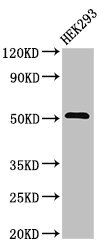
Western Blot
Positive WB detected in: HEK293 whole cell lysate
All lanes: TNFRSF10A antibody at 3.2μg/ml
Secondary
Goat polyclonal to rabbit IgG at 1/50000 dilution
Predicted band size: 51 kDa
Observed band size: 51 kDa
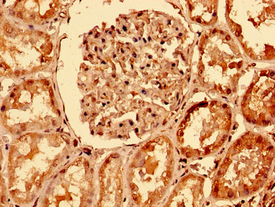
IHC image of CSB-PA023964LA01HU diluted at 1:400 and staining in paraffin-embedded human kidney tissue performed on a Leica BondTM system. After dewaxing and hydration, antigen retrieval was mediated by high pressure in a citrate buffer (pH 6.0). Section was blocked with 10% normal goat serum 30min at RT. Then primary antibody (1% BSA) was incubated at 4°C overnight. The primary is detected by a biotinylated secondary antibody and visualized using an HRP conjugated SP system.
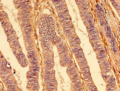
IHC image of CSB-PA023964LA01HU diluted at 1:400 and staining in paraffin-embedded human colon cancer performed on a Leica BondTM system. After dewaxing and hydration, antigen retrieval was mediated by high pressure in a citrate buffer (pH 6.0). Section was blocked with 10% normal goat serum 30min at RT. Then primary antibody (1% BSA) was incubated at 4°C overnight. The primary is detected by a biotinylated secondary antibody and visualized using an HRP conjugated SP system.
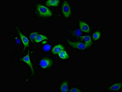
Immunofluorescent analysis of A549 cells using CSB-PA023964LA01HU at dilution of 1:100 and Alexa Fluor 488-congugated AffiniPure Goat Anti-Rabbit IgG(H+L)
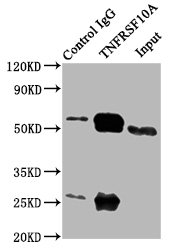
Immunoprecipitating TNFRSF10A in 293 whole cell lysate
Lane 1: Rabbit control IgG (1μg) instead of CSB-PA023964LA01HU in 293 whole cell lysate.
For western blotting, a HRP-conjugated Protein G antibody was used as the secondary antibody (1/2000)
Lane 2: CSB-PA023964LA01HU (6μg) + 293 whole cell lysate (500μg)
Lane 3: 293 whole cell lysate (10μg)
The following TNFRSF10A reagents supplied by CUSABIO are manufactured under a strict quality control system. Multiple applications have been validated and solid technical support is offered.
TNFRSF10A Antibodies for Homo sapiens (Human)
| Code | Product Name | Species Reactivity | Application |
|---|---|---|---|
| CSB-PA199651 | TNFRSF10A Antibody | Human | ELISA,WB |
| CSB-PA002191 | TNFRSF10A Antibody | Human,Monkey | WB, IF, ELISA |
| CSB-PA023964LA01HU | TNFRSF10A Antibody | Human | ELISA, WB, IHC, IF, IP |
| CSB-PA023964LB01HU | TNFRSF10A Antibody, HRP conjugated | Human | ELISA |
| CSB-PA023964LC01HU | TNFRSF10A Antibody, FITC conjugated | Human | |
| CSB-PA023964LD01HU | TNFRSF10A Antibody, Biotin conjugated | Human | ELISA |
TNFRSF10A Proteins for Homo sapiens (Human)
| Code | Product Name | Source |
|---|---|---|
| CSB-CF023964HU | Recombinant Human Tumor necrosis factor receptor superfamily member 10A (TNFRSF10A) | in vitro E.coli expression system |
| CSB-YP023964HU1 CSB-EP023964HU1 CSB-BP023964HU1 CSB-EP023964HU1-B |
Recombinant Human Tumor necrosis factor receptor superfamily member 10A (TNFRSF10A), partial | Yeast E.coli Baculovirus In Vivo Biotinylation in E.coli |

