Call us
301-363-4651 (Available 9 a.m. to 5 p.m. CST from Monday to Friday)
Mesothelin, also known as MSLN, is a cell surface glycoprotein encoded by the MSLN gene. The MSLN gene encodes a precursor protein that is enzymatically decomposed into two products: megakaryocyte-potentiating factor (MPF) and mesothelin. MPF is a cytokine that stimulates the formation of bone marrow megakaryocyte colonies. Mesothelin is a protein anchored to the cell surface by glycosylphosphatidylinositol.
Mesothelin is a differentiated antigen found in normal mesotheliocytes. It is rarely expressed in normal tissues, but it is highly expressed in mesothelioma, lung cancer, pancreatic cancer, breast cancer, ovarian cancer and other tumors [1]. Therefore, mesothelioma may become an important target for cancer treatment.
1. The Discovery of Mesothelin
2. Structural Characteristics of Mesothelin
This protein was first identified by its reactivity with monoclonal antibody K1 [2]. Mesothelin is specifically recognized by monoclonal antibody K1. Chang et al. extracted complementary DNA from a gene library of a Hela cell line that reacts with monoclonal antibody K1, and cloned the mesothelin gene, which was named mesothelin. It is named mesothelin because the membrane-bound protein produced by this gene is mainly located on the surface of normal mesothelial cells.
The mesothelin gene is located on chromosome 16 p13.3. Its gene is 8 kb in length and its cDNA size is 213 bp. It contains 1884 bp open reading frame and 17 exons encoding 628 amino acids.
The MSLN gene encodes a 71 KD precursor protein. The precursor protein is hydrolyzed by furin protease into two parts: a soluble 31 kD N-terminal protein called megakaryocyte-potentiating factor (MPF) and a 40 kD membrane-bound fragment called MSLN [3].
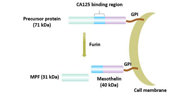
Figure 1 Maturation process of mesothelitin
At present, there are at least 3 isomers of mesothatin. Membrane-bound mesothelin is the most common variant of mesothelin; mesothelin V-1 has an additional 8 amino acids and it is also a membrane-bound form; mesothelin V-2 has a modified COOH end. It is a mesothelin-related peptide (SMRP) that is shed from the cell surface after hydrolysis by protease [4].
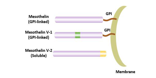
Figure 2 Variant of mesothelin
The soluble MSLN-related protein is generally 42-45 kDa in size, and its NH2-terminal amino acid sequence is identical to membrane-bound mesothelin. The production of SMRP could be related to abnormal splicing, which results in a secreted form or its cleavage from the membrane by the TNFα-converting enzyme ADAM17.
SMRP can be detected in the blood of some mesothelin-positive tumor patients.
The detection of blood mesothelin can be used in the diagnosis of some diseases.
Mesothelin is expressed in mesothelial cells of the normal pleura, pericardium and peritoneum, and it has the lowest expression on the surface of epithelial cells of the trachea, ovaries, testes, tonsils and fallopian tubes. In addition, as a cell surface antigen associated with tumor invasion, it is also highly expressed in a variety of tumor tissues, mainly including mesothelioma, ovarian and pancreatic adenocarcinoma.
MSLN was found to be overexpressed in a variety of cancers.
Currently, the overexpression of mesothelin has been observed in mesothelioma, ovarian cancer, lung cancer, esophageal cancer, pancreatic cancer [5], gastric cancer, cholangiocarcinoma, endometrial cancer, thymic cancer, colon cancer and breast cancer.
The expression frequency and distribution of MSLN vary with tumor subtypes. MSLN was found to be expressed in 90% of epithelioid malignant pleural mesothelioma, 69% of lung adenocarcinoma, 60% of breast cancer and 46% of esophageal cancer [6].
In cancer cells, MSLN expression may be in the lumen/membrane or cytoplasm. The expression location of MSLN in different types of tumors is also different. In mesothelioma tumors, MSLN expression was uniformly distributed on the cell surface.
In lung adenocarcinoma, MSLN is expressed in both cytoplasm and cell surface.
Cytoplasmic expression was more common than membrane expression in gastric cancer.
MSLN is also expressed in solid tumors such as thyroid, kidney and synovial sarcoma tumors.
Researchers have studied MSLN knockout mice and found that these knockout mice showed normal development, reproduction, and blood cell counts [7]. This indicates that mesothelin is not required for normal growth and reproduction of mice. Because of this, the biological function of mesothelin has not been known so far.
Studies have shown that mesothelin may play a role in adhesion. With the deepening of research on tumor, mesothelin has been found to play an important role in the development of various tumors. Overexpression of MSLN activates NF-κB, MAPK and PI3K pathways and induces apoptosis or promotes cell proliferation, migration and metastasis by inducing activation and expression of MMP7 and MMP9.
Abnormal expression of MSLN plays an important role in tumor cell proliferation, adhesion and drug resistance.
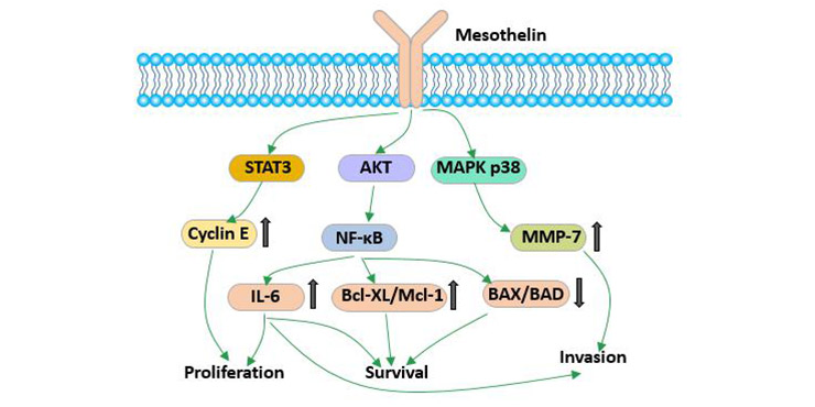
Figure 3 The biological function of mesothelin
In the study of pancreatic cancer, it was found that mesothelin can promote tumor cell proliferation. Silencing the mesothelin gene in pancreatic cancer tumor cells can inhibit proliferation.
Mesothelin can inhibit cell cycle by slowing down cell entry into S phase [8].
Excessive expression of mesothelin can continuously activate the NF- kappaB signaling pathway and produce high levels of IL-6, thereby activating transcriptional protein Stat3, resulting in increased expressions of cyclin E and cyclin-dependent kinase 2 (CDK2). This eventually accelerated the cell transition from G1 to S.
Studies have shown that mouse embryonic fibroblasts 3T3 transfected with mesothelin are more strongly attached to the culture dish and are more difficult to remove from the cell surface than untransfected cells, suggesting that mesothelin may be involved in cell adhesion.
In the study of ovarian cancer, Rump et al. [9] showed that the high affinity interaction between MSLN and CA125 resulted in heterogeneous adhesion between cells and promoted the metastasis of ovarian cancer cell lines. The expression of MSLN and the increase of serum SMRP level are related to the progress of tumor, the increase of stage and the decrease of overall survival rate.
Mesothelin can increase the tolerance of cancer cells to drugs [10] [11].
Studies have shown that mesothelin expression is different in chemotherapeutic sensitive and drug-resistant patients with ovarian cancer, and the expression of mesothelin is significantly elevated in drug-resistant patients. After down-regulating mesothelin expression, the chemosensitivity of ovarian cancer resistant cell lines was also significantly improved, which reversed cell resistance to some extent.
Studies have shown that mesothatin may block taxol-induced tumor cell apoptosis by simultaneously activating PI3K/AKT and MAPK/ERK pathways. This is one of the causes of cell resistance induced by mesothelin.
Mesothelin gene may be regulated by molecules of Wnt signaling pathway [12]. In tumors with sustained activation of Wnt signaling pathways, such as ovarian cancer and pancreatic cancer, the expression of mesothelin increased.
Mesothelin is a protein with strong immunogenicity. In addition, it has less expression in normal tissues and is highly expressed in malignant tumor cells. These characteristics make mesothelin a target for anti-tumor vaccines and cancer treatment.
CRS-207 is a tumor vaccine with listeria as the carrier. Its mechanism of action is:
For invading bacteria, macrophages phagocytose, internalize and process them, present mesothelin antigen to T cells, release lymphokines, activate killer T cells to target mesothelin, and finally cause apoptosis of tumor cells expressing mesothelin.
Soluble mesothelin can be detected in serum of patients with solid tumors, which makes it a valuable diagnostic tool for tumors expressing mesothelin. Plasma soluble mesocertin was detected by sandwich ELISA. Soluble mesothelin itself lacks sensitivity and specificity, but combined detection with CA125 can improve diagnostic efficiency [13]. Mesothelin combined with other ovarian tumor markers can be used for screening for ovarian cancer.
Given that mesothelin is highly expressed in a variety of human malignant cells and is rarely expressed in normal tissues, mesothelin is expected to be a target for specific treatment of tumors. Various immunotherapeutic strategies have been designed for MSLN, including the design of drugs based on antibody therapy and CAR-T. The MSLN targeted immunotherapy reported to date has good safety [14] [15].
Antibody-based drugs can target and kill tumor cells via a variety of ways. It mainly including antibody neutralization, antibody-dependent cell-mediated cytotoxicity (ADCC), antibody-dependent cell-mediated phagocytosis (ADCP) or antibodies conjugated with effector molecules (toxins or inhibitors), which mediate apoptosis or suppress cell proliferation.
Amatuximab (MORAb-009) is a chimeric antibody composed of single strand variable region fragments of anti-mesothatin antibody SS1 and human IgG1 and κ constant regions. It kills tumor cells through antibody-dependent cell-mediated cytotoxicity (ADCC) and blocks the interaction between mesothatin and CA125 [16].
Anetumab ravtansine (BAY94-9343) is an antibody-conjugated drug consisting of an anti-mesothelin antibody and a maytansin derivative DM4 (tubulin polymerase inhibitor) [17]. BAY94-9343 binds to mesothelin on the surface of tumor cells, then it can be internalized into lysosomes and release DM4 to kill tumor cells
DMOT4039A is an antibody-conjugated drug obtained by linking a humanized IgG1 anti-mesothelin monoclonal antibody h7D9.v3 and an antimitotic agent monomethyl auristatin E (MMAE) via a dipeptide. DMOT4039A mainly enters lysosomes by clathrin-mediated endocytosis and releases MMAE. Subsequently, MMAE binds to cytoplasmic tubulin to block the G2/M phase of the cell cycle, arresting cell growth and inducing apoptosis.
MDX-1204 is an antibody-conjugated drug conjugated with a fully human mesothelin antibody (MDX-1383) and an alkylating agent duocarmycin (MED2460) [18]. The binding of MDX-1204 to mesothelin blocks its interaction with CA125. After binding to mesothelin, MDX-1204 is internalized by the cell and releases the small molecule drug MED2460. MED2460 binds to the minor groove of DNA, causing irreversible alkylation of DNA, which destroys the nucleic acid structure and ultimately leads to cell death.
SS1P is a recombinant immunotoxin, which is formed by fusing the variable region fragment of mouse anti-mesodermal antibody with pseudomonas exotoxin 38 (PE38) [19]. It is currently undergoing clinical evaluation in patients with tumors that express mesothelin. Many drugs based on the MSLN antibody SS1 or other modified and humanized versions have been developed for targeted therapies.
Chimeric antigen receptor T cells (CAR) are usually composed of extracellular antigen recognition domains (which are usually derived from single chain variable fragments (scFv) of antibodies), transmembrane domains and intracellular domains that transmit activation signals of T cells.
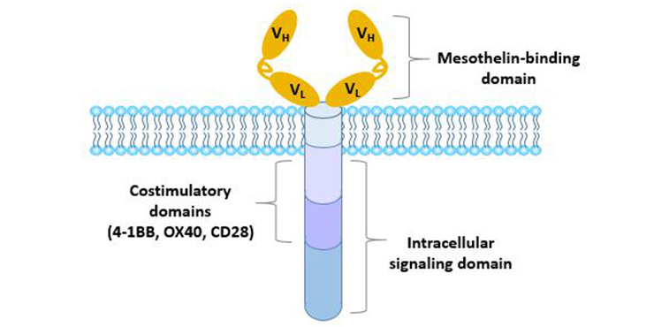
Figure 4 Structure of chimeric antigen receptor
The first generation of CAR contains only one intracellular signal domain, usually the CD3 ζ chain. This is sufficient to initiate T cell activation, with the disadvantage of producing only short-term proliferative activity and low levels of cytotoxicity.
The second generation of CAR greatly improves its efficacy by adding another co-stimulator (CD28, 4-1BB or OX40).
The third generation CAR contains two costimulatory domains (CD28, 4-1BB, TLR2 or DAP10) and a hinge domain, which is superior in cell proliferation, cytotoxicity, persistence and tumor suppressive effects.
The fourth generation of CAR can secrete cytokines or other effector molecules such as IL-12, IL-15, IL-7 or CCL19 to regulate the immune microenvironment.
More details about CAR-T are available in this article: CAR-T Cell Therapy- An Useful Treatment To Cancer.
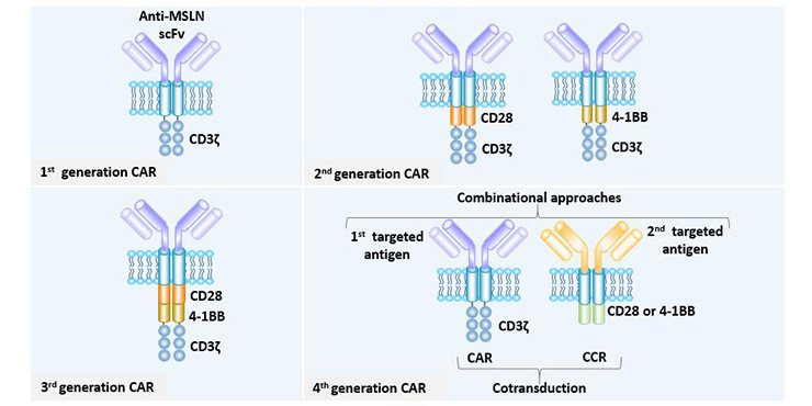
Figure 5 Different generations of the MSLN CAR
Mesothelin CAR-T cell therapy has achieved encouraging results in mouse models of mesothelioma, ovarian cancer, and lung cancer, and some clinical trials have been approved and are currently underway [20] [21].
Improving the efficiency of CAR-T cells is one of the main problems in CAR-T therapy. This mainly needs to be solved from the following aspects:
Promote CAR-T cell infiltration;
Enhance the functional persistence of CAR-T cells;
Enhancing CAR-T cells to overcome the inhibition signals;
Improving safety by preventing on-target/off-tumor toxicity.
Designing MSLN CAR-T cells to overexpress the chemokine receptor CCR2B has been shown to enhance the transport of T cells to solid tumors. Overexpression of CCR2B significantly increases the therapeutic efficiency of systemic administration.
References
[1] Paolo B, Sara C. Amatuximab and novel agents targeting mesothelin for solid tumors [J]. OncoTargets and Therapy, 2017, Volume 10: 5337-5353.
[2] Chang K. Characterization of the antigen (CAK1) recognized by monoclonal antibody K1 present on ovarian cancers and normal mesothelium [J]. Cancer Res, 1992, 52(1): 181-186.
[3] Manzanares M, Campbell D J W, Maldonado G T, et al. Overexpression of periostin and distinct mesothelin forms predict malignant progression in a rat cholangiocarcinoma model [J]. Hepatol Commun, 2018, 2(2): 155-172.
[4] Burt B M, Lee H S, Rosen V L D, et al. Soluble Mesothelin-Related Peptides to Monitor Recurrence After Resection of Pleural Mesothelioma [J]. Annals of Thoracic Surgery, 2017, 104(5): 1679.
[5] Argani P. Mesothelin is overexpressed in the vast majority of ductal adenocarcinomas of the pancreas: Identification of a new pancreatic cancer marker by serial analysis of gene expression (SAGE) [J]. Clin Cancer Res, 2001, 7.
[6] Morello A, Sadelain M, Adusumilli P S. Mesothelin-Targeted CARs: Driving T cells to Solid Tumors [J]. Cancer Discovery, 2015, 6(2).
[7] Bera T K, Pastan I. Mesothelin Is Not Required for Normal Mouse Development or Reproduction [J]. Molecular and Cellular Biology, 2000, 20(8): 2902-2906.
[8] Guadalupe A M, Beate P, Calderón-Aranda Emma S, et al. Biomarkers for Predicting Malignant Pleural Mesothelioma in a Mexican Population [J]. International Journal of Medical Sciences, 2018, 15(9): 883-891.
[9] Rump A, Morikawa Y, Tanaka M, et al. Binding of Ovarian Cancer Antigen CA125/MUC16 to Mesothelin Mediates Cell Adhesion [J]. Journal of Biological Chemistry, 2004, 279(10): 9190-9198.
[10] Bharadwaj U, Marin-Muller C, Li M, et al. Mesothelin confers pancreatic cancer cell resistance to TNF-α-induced apoptosis through Akt/PI3K/NF-κB activation and IL-6/Mcl-1 overexpression [J]. Molecular Cancer, 2011, 10(1): 106.
[11] Cheng W F, Huang C Y, Chang M C, et al. High mesothelin correlates with chemoresistance and poor survival in epithelial ovarian carcinoma [J]. BRITISH JOURNAL OF CANCER, 2009, 100(7): 1144-1153.
[12] Moon R T, Prieve M G. Stromelysin-1 and mesothelin are differentially regulated by Wnt-5a and Wnt-1 in C57mg mouse mammary epithelial cells [J]. BMC Developmental Biology, 2003, 3(1): 1-10.
[13] Bast R C. Status of tumor markers in ovarian cancer screening [J]. Journal of clinical oncology: official journal of the American Society of Clinical Oncology, 2003, 21(10 Suppl): 200s.
[14] Villena-Vargas J, Adusumilli P S. Mesothelin-targeted immunotherapies for malignant pleural mesothelioma [J]. Annals of Cardiothoracic Surgery, 2012, 1(4): 466.
[15] Pastan I, Hassan R. Discovery of Mesothelin and Exploiting It as a Target for Immunotherapy [J]. Cancer Research, 2014, 74(11): 2907-2912.
[16] Hassan R, Ebel W, Routhier E L, et al. Preclinical evaluation of MORAb-009, a chimeric antibody targeting tumor-associated mesothelin [J]. Cancer Immunity, 2007, 7: 20.
[17] Grosso F, Scagliotti G V. Systemic treatment of malignant pleural mesothelioma [J]. Future Oncology, 2012, 8(3): 293-305.
[18] Pogue S, Chen X, Alexander S, et al. Fully human antimesothelin antibody drug conjugate demonstrates antitumor effects in human lung cancer models [J]. Clinical Cancer Research, 2008, 14(15 Supplement): A13-A13.
[19] Kolyvas E, Rudloff M, Poruchynsky M, et al. Mesothelin-targeted immunotoxin RG7787 has synergistic anti-tumor activity when combined with taxanes [J]. Oncotarget, 2017, 8(6): 9189.
[20] Beatty GL, Haas AR, Maus MV, et al. Mesothelin-Specific Chimeric Antigen Receptor mRNA-Engineered T Cells Induce Antitumor Activity in Solid Malignancies [J]. Cancer Immunology Research, 2015, 3(2): 217.
[21] Adusumilli P S, Cherkassky L, Villena-Vargas J, et al. Regional delivery of mesothelin-targeted CAR T cell therapy generates potent and long-lasting CD4-dependent tumor immunity [J]. Science Translational Medicine, 2014, 6(261): 261ra151-261ra151.


Comments
Leave a Comment