Call us
301-363-4651 (Available 9 a.m. to 5 p.m. CST from Monday to Friday)
Calpain-2, encoded by CAPN2 and commonly known as m-calpain, is a calcium-dependent cysteine protease activated by moderate Ca²+ concentrations. It cleaves cytoskeletal proteins (e.g., actin) and signaling molecules, regulating cell migration, proliferation, and apoptosis, with key roles in PI3K/Akt and MAPK pathways.
Dysregulation of Calpain-2 links to breast/colorectal cancer invasion via extracellular matrix remodeling and Alzheimer’s disease via tau protein cleavage. Selective inhibitors like Calpeptin are in preclinical/early clinical stages, targeting its catalytic domain to reduce pathological activity.
Recombinant Human Calpain-2 catalytic subunit (CAPN2) (CSB-EP004496HU)
Validated Data

(Tris-Glycine gel) Discontinuous SDS-PAGE (reduced) with 5% enrichment gel and 15% separation gel.
CAPN2 Antibody (CSB-PA004496LA01HU)
Validated Data
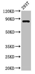
Western Blot
Positive WB detected in: 293T whole cell lysate
All lanes: CAPN2 antibody at 3.4µg/ml
Secondary
Goat polyclonal to rabbit IgG at 1/50000 dilution
Predicted band size: 80, 72 kDa
Observed band size: 80 kDa
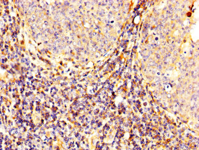
IHC image of CSB-PA004496LA01HU diluted at 1:300 and staining in paraffin-embedded human tonsil tissue performed on a Leica BondTM system. After dewaxing and hydration, antigen retrieval was mediated by high pressure in a citrate buffer (pH 6.0). Section was blocked with 10% normal goat serum 30min at RT. Then primary antibody (1% BSA) was incubated at 4°C overnight. The primary is detected by a biotinylated secondary antibody and visualized using an HRP conjugated SP system.
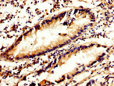
IHC image of CSB-PA004496LA01HU diluted at 1:300 and staining in paraffin-embedded human appendix tissue performed on a Leica BondTM system. After dewaxing and hydration, antigen retrieval was mediated by high pressure in a citrate buffer (pH 6.0). Section was blocked with 10% normal goat serum 30min at RT. Then primary antibody (1% BSA) was incubated at 4°C overnight. The primary is detected by a biotinylated secondary antibody and visualized using an HRP conjugated SP system.
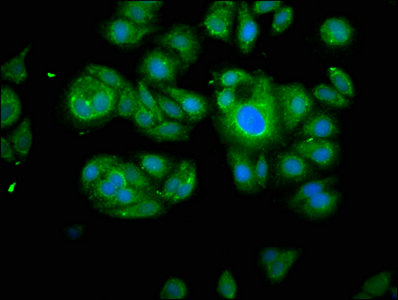
Immunofluorescence staining of HepG2 cells with CSB-PA004496LA01HU at 1:100, counter-stained with DAPI. The cells were fixed in 4% formaldehyde, permeabilized using 0.2% Triton X-100 and blocked in 10% normal Goat Serum. The cells were then incubated with the antibody overnight at 4°C. The secondary antibody was Alexa Fluor 488-congugated AffiniPure Goat Anti-Rabbit IgG(H+L).
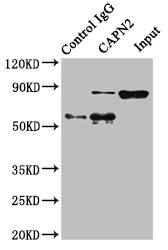
Immunoprecipitating CAPN2 in Hela whole cell lysate
Lane 1: Rabbit control IgG instead of CSB-PA004496LA01HU in Hela whole cell lysate. For western blotting, a HRP-conjugated Protein G antibody was used as the secondary antibody (1/2000)
Lane 2: CSB-PA004496LA01HU (8µg) + Hela whole cell lysate (500µg)
Lane 3: Hela whole cell lysate (10µg)
Human calpain 2, (m/II) large subunit (CAPN2) ELISA kit (CSB-E17822h)
Validated Data
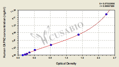
Code: CSB-E17822h
Size: 96T,5×96T,10×96T
Sensitivity: 0.039 ng/mL
Detection Range: 0.156 ng/mL-10 ng/mL
These standard curves are provided for demonstration only. A standard curve should be generated for each set of samples assayed.
The following CAPN2 reagents supplied by CUSABIO are manufactured under a strict quality control system. Multiple applications have been validated and solid technical support is offered.
CAPN2 Antibodies for Homo sapiens (Human)
| Code | Product Name | Species Reactivity | Application |
|---|---|---|---|
| CSB-PA004496LA01HU | CAPN2 Antibody | Human | ELISA, WB, IHC, IF, IP |
| CSB-PA004496GA01HU | CAPN2 Antibody | Human,Mouse,Rat | ELISA,WB,IHC |
| CSB-PA060228 | CAPN2 Antibody | Human,Mouse,Rat | IHC, ELISA |
| CSB-PA257142 | CAPN2 Antibody | Human,Mouse,Rat | ELISA,WB |
| CSB-PA247305 | CAPN2 Antibody | Human,Mouse,Rat | ELISA,WB |
| CSB-RA899086A0HU | CAPN2 Recombinant Monoclonal Antibody | Human | ELISA, WB |
CAPN2 Antibodies for Pig
| Code | Product Name | Species Reactivity | Application |
|---|---|---|---|
| CSB-PA004496LA01PI | CAPN2 Antibody | Pig | ELISA |
CAPN2 Proteins for Homo sapiens (Human)
| Code | Product Name | Source |
|---|---|---|
| CSB-YP004496HU CSB-BP004496HU CSB-MP004496HU CSB-EP004496HU-B |
Recombinant Human Calpain-2 catalytic subunit (CAPN2) | Yeast Baculovirus Mammalian cell In Vivo Biotinylation in E.coli |
| CSB-EP004496HU | Recombinant Human Calpain-2 catalytic subunit (CAPN2) | E.coli |
CAPN2 Proteins for Sus scrofa (Pig)
| Code | Product Name | Source |
|---|---|---|
| CSB-YP004496PI CSB-BP004496PI CSB-MP004496PI CSB-EP004496PI-B |
Recombinant Pig Calpain-2 catalytic subunit (CAPN2) | Yeast Baculovirus Mammalian cell In Vivo Biotinylation in E.coli |
| CSB-EP004496PI | Recombinant Pig Calpain-2 catalytic subunit (CAPN2) | E.coli |
CAPN2 Proteins for Oryctolagus cuniculus (Rabbit)
| Code | Product Name | Source |
|---|---|---|
| CSB-YP004496RB CSB-EP004496RB CSB-BP004496RB CSB-MP004496RB CSB-EP004496RB-B |
Recombinant Rabbit Calpain-2 catalytic subunit (CAPN2) | Yeast E.coli Baculovirus Mammalian cell In Vivo Biotinylation in E.coli |
CAPN2 Proteins for Mus musculus (Mouse)
| Code | Product Name | Source |
|---|---|---|
| CSB-YP004496MO CSB-EP004496MO CSB-BP004496MO CSB-MP004496MO CSB-EP004496MO-B |
Recombinant Mouse Calpain-2 catalytic subunit (Capn2) | Yeast E.coli Baculovirus Mammalian cell In Vivo Biotinylation in E.coli |
CAPN2 Proteins for Bos taurus (Bovine)
| Code | Product Name | Source |
|---|---|---|
| CSB-YP631683BO CSB-EP631683BO CSB-BP631683BO CSB-MP631683BO CSB-EP631683BO-B |
Recombinant Bovine Calpain-2 catalytic subunit (CAPN2), partial | Yeast E.coli Baculovirus Mammalian cell In Vivo Biotinylation in E.coli |
CAPN2 Proteins for Gallus gallus (Chicken)
| Code | Product Name | Source |
|---|---|---|
| CSB-YP849716CH CSB-EP849716CH CSB-BP849716CH CSB-MP849716CH CSB-EP849716CH-B |
Recombinant Chicken Calpain-2 catalytic subunit (CAPN2), partial | Yeast E.coli Baculovirus Mammalian cell In Vivo Biotinylation in E.coli |
CAPN2 Proteins for Macaca fascicularis (Crab-eating macaque) (Cynomolgus monkey)
| Code | Product Name | Source |
|---|---|---|
| CSB-YP867083MOV CSB-EP867083MOV CSB-BP867083MOV CSB-MP867083MOV CSB-EP867083MOV-B |
Recombinant Macaca fascicularis Calpain-2 catalytic subunit (CAPN2), partial | Yeast E.coli Baculovirus Mammalian cell In Vivo Biotinylation in E.coli |
CAPN2 ELISA Kit for Homo sapiens (Human)
| Code | Product Name | Sample Types | Sensitivity |
|---|---|---|---|
| CSB-E17822h | Human calpain 2, (m/II) large subunit (CAPN2) ELISA kit | serum, plasma, cell lysates | 0.039 ng/mL |

