Recombinant Mouse Macrophage colony-stimulating factor 1 protein (Csf1), partial (Active)

In Stock
Code CSB-AP003461MO
Abbreviation Recombinant Mouse Csf1 protein, partial (Active)
MSDS
Size $354
Order now
Image
Have Questions? Leave a Message or Start an on-line Chat

Product Details

Purity
>95% as determined by SDS-PAGE.
Endotoxin
Less than 1.0 EU/μg as determined by LAL method.
Activity
Fully biologically active when compared to standard. The ED50 as determined by a cell proliferation assay using murine M-NFS-60 cells is less than 2 ng/ml, corresponding to a specific activity of >5.0x10⁵ IU/mg.
Target Names
Uniprot No.
Research Area
Immunology
Alternative Names
Csf1; CsfmMacrophage colony-stimulating factor 1; CSF-1; MCSF) [Cleaved into: Processed macrophage colony-stimulating factor 1]
Species
Mus musculus (Mouse)
Source
E.Coli
Expression Region
33-262aa
Complete Sequence
KEVSEHCSHM IGNGHLKVLQ QLIDSQMETS CQIAFEFVDQ EQLDDPVCYL KKAFFLVQDI IDETMRFKDN TPNANATERL QELSNNLNSC FTKDYEEQNK ACVRTFHETP LQLLEKIKNF FNETKNLLEK DWNIFTKNCN NSFAKCSSRD VVTKPDCNCL YPKATPSSDP ASASPHQPPA PSMAPLAGLA WDDSQRTEGS SLLPSELPLR IEDPGSAKQR PPRSTCQTLE
Mol. Weight
26 kDa
Protein Length
Partial
Tag Info
Tag-Free
Form
Lyophilized powder
Buffer
Lyophilized from a 0.2 µm filtered 20 mM Tris, 500 mM NaCl, pH 7.4
Reconstitution
We recommend that this vial be briefly centrifuged prior to opening to bring the contents to the bottom. Please reconstitute protein in deionized sterile water to a concentration of 0.1-1.0 mg/mL.We recommend to add 5-50% of glycerol (final concentration) and aliquot for long-term storage at -20°C/-80°C. Our default final concentration of glycerol is 50%. Customers could use it as reference.
Troubleshooting and FAQs
Storage Condition
Store at -20°C/-80°C upon receipt, aliquoting is necessary for mutiple use. Avoid repeated freeze-thaw cycles.
Shelf Life
The shelf life is related to many factors, storage state, buffer ingredients, storage temperature and the stability of the protein itself.
Generally, the shelf life of liquid form is 6 months at -20°C/-80°C. The shelf life of lyophilized form is 12 months at -20°C/-80°C.
Lead Time
5-10 business days
Notes
Repeated freezing and thawing is not recommended. Store working aliquots at 4°C for up to one week.
Datasheet & COA
Please contact us to get it.
Description

Recombinant Mouse Macrophage colony-stimulating factor 1 protein (Csf1) comes from an E. coli expression system and covers amino acids 33-262 of the protein. This tag-free version shows purity levels above 95% when checked by SDS-PAGE analysis. The protein appears to be fully biologically active, demonstrating an ED50 of less than 2 ng/ml in murine M-NFS-60 cell proliferation assays. This translates to a specific activity greater than 5.0 × 10^5 IU/mg. Endotoxin levels stay below 1.0 EU/µg, as measured by the LAL method.

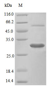
Macrophage colony-stimulating factor 1 (Csf1) seems to play a critical role in how mononuclear phagocytes are produced, differentiate, and function. The immune system likely depends on this protein for processes like macrophage activation and survival. Research in immunology, hematopoiesis, and cellular biology often turns to Csf1 as a key tool for understanding macrophage-related pathways and how cells interact with each other.

Potential Applications

Note: The applications listed below are based on what we know about this protein's biological functions, published research, and experience from experts in the field. However, we haven't fully tested all of these applications ourselves yet. We'd recommend running some preliminary tests first to make sure they work for your specific research goals.

1. Cell Proliferation and Viability Assays for Macrophage Research

This recombinant mouse Csf1 protein is confirmed to be biologically active (ED₅₀ < 2 ng/ml in murine M-NFS-60 cells) and can be used to stimulate macrophage proliferation and survival. However, since the protein is a partial sequence (33-262aa) and M-NFS-60 cells are typically used for G-CSF (Csf3) assays, researchers should validate its activity in more relevant macrophage-specific cell lines (e.g., RAW264.7 or bone marrow-derived macrophages) to ensure physiological relevance. The high specific activity (>5.0×10⁵ IU/mg) and low endotoxin levels support reliable dose-response studies, but results may not fully represent native Csf1 function due to the truncated form.

2. Biochemical Characterization and Receptor Binding Studies

The tag-free design and high purity (>95%) make this protein suitable for biochemical studies of Csf1-receptor interactions. However, the partial sequence may lack full receptor-binding domains present in the full-length protein, potentially affecting binding kinetics and affinity measurements. Researchers should interpret data from surface plasmon resonance or competitive binding assays with caution and validate key findings with full-length Csf1 when possible.

3. Antibody Development and Validation

This biologically active Csf1 can serve as an immunogen for antibody development, but the partial sequence may generate antibodies that do not recognize full-length Csf1. Antibodies should be validated against native, full-length Csf1 in biological samples to ensure recognition of all relevant epitopes. The high purity and defined sequence are advantageous for standardization, but functional neutralization assays may require confirmation with full-length protein.

4. Signal Transduction Pathway Analysis

The protein is appropriate for studying Csf1 signaling pathways due to its confirmed activity, but the E. coli expression system lacks mammalian post-translational modifications, which could alter signaling kinetics or amplitude. Researchers should verify that phosphorylation events (e.g., MAPK, PI3K pathways) mirror those induced by native Csf1, especially in primary cells.

5. Preclinical Model Development and Validation

This recombinant Csf1 can be used in preclinical models, but the partial sequence may not fully recapitulate native Csf1 function in complex systems. While low endotoxin levels and biological activity support reproducibility, researchers should validate key outcomes (e.g., macrophage differentiation) with full-length Csf1 to ensure translational relevance.

Final Recommendation & Action Plan

This E. coli-expressed partial mouse Csf1 protein (33-262aa) has validated biological activity and is suitable for initial studies, but its truncated nature requires careful validation for each application. Prioritize confirming activity in macrophage-specific cell lines beyond M-NFS-60 to ensure relevance. For binding and signaling studies, compare results with full-length Csf1 to account for potential domain deficiencies. When developing antibodies, test cross-reactivity with native protein. While the high purity and low endotoxin levels make it useful for controlled assays, critical findings should be verified with full-length Csf1 to ensure biological accuracy. Always include appropriate controls and dose-response curves to mitigate limitations associated with the partial sequence.

Customer Reviews and Q&A

 Customer Reviews

There are currently no reviews for this product.

Submit a Review here

Target Background

Function
Cytokine that plays an essential role in the regulation of survival, proliferation and differentiation of hematopoietic precursor cells, especially mononuclear phagocytes, such as macrophages and monocytes. Promotes the release of proinflammatory chemokines, and thereby plays an important role in innate immunity and in inflammatory processes. Plays an important role in the regulation of osteoclast proliferation and differentiation, the regulation of bone resorption, and is required for normal bone development. Required for normal male and female fertility. Promotes reorganization of the actin cytoskeleton, regulates formation of membrane ruffles, cell adhesion and cell migration. Plays a role in lipoprotein clearance.
Gene References into Functions
  1. In glioblastoma, colony-stimulating factor-1 and angiocrine IL-6 induce robust arginase-1 expression and macrophage alternative activation, mediated through peroxisome proliferator-activated receptor-gamma-dependent transcriptional activation of hypoxia-inducible factor-2alpha. PMID: 29422647
  2. M-CSF serves as an intermediate signal, thus inducing a vital decrease in the NPR2 levels in cumulus cells, and regulates the process of LH-induced resumption of meiosis. PMID: 28978329
  3. Findings revealed that stress-induced elevations in neuronal CSF1 provokes microglia-mediated neuronal remodeling in layer 1 medial prefrontal cortex, contributing to synaptic deficits and development of anxiety- and depressive-like behavior. Moreover, chronic stress exposure elicited divergent neuroimmune responses in male and female mice, demonstrating sex-dependent differences in neuron-microglia interactions. PMID: 28697890
  4. PLEKHO2-deficient bone marrow-derived macrophages displayed increased apoptotic cell death in the absence of Macrophage-colony stimulating factor, although PLEKHO2 deficiency did not affect macrophage differentiation and proliferation. PMID: 28627369
  5. lymphatic endothelial cells cause bone destruction in vivo in mice by secreting M-CSF, which promotes osteoclasts formation and activation. PMID: 28052488
  6. address which CSF-1-activated pathways are involved in transmitting the lineage-instructive signal in primary bone marrow-derived GM progenitors. PMID: 28159742
  7. study concludes that M-CSF is critical to host defenses against bacterial pneumonia by mediating survival and antimicrobial functions of mononuclear phagocytes in the lungs and liver. PMID: 27183631
  8. study, therefore, provided insights into the sequence-structure-function relationships of the M-CSF/c-FMS interaction and of ligand/receptor tyrosine kinase interactions in general. PMID: 28655719
  9. Therefore, our findings indicate that CSF1 signaling is oncogenic during gliomagenesis through a mechanism distinct from modulating glioma-associated microglia/macrophage polarization status PMID: 27013192
  10. study concludes that Langerhans cells require IL-34 when residing in fully differentiated and anatomically intact skin epidermis, but rely on neutrophil-derived CSF1 during inflamma PMID: 26634935
  11. Proteomic Analysis Reveals Distinct Metabolic Differences Between Granulocyte-Macrophage Colony Stimulating Factor (GM-CSF) and Macrophage Colony Stimulating Factor (M-CSF) Grown Macrophages Derived from Murine Bone Marrow Cells PMID: 26229149
  12. Hematopoietic cells can be induced by M-CSF to dedifferentiate to multipotent stem cells. PMID: 26529564
  13. M-CSF promotes macrophagic over granulocytic differentiation by inducing ERK activation but also PKCd expression, which in turn, down-regulates Fli-1 expression and prevents granulocytic differentiation. PMID: 26336156
  14. Results identify CSF-1-activated macrophages as crucial mediators of detrimental Schwann cell dedifferentiation in Cx32-deficient mice PMID: 25628221
  15. Ceramide production in M-CSF-deprived macrophages arises from a combination of ASMase activity and de novo synthesis. PMID: 26253821
  16. CSF-1 did not rescue the growth and lung defects associated with hyperoxia in this model; however, an increase in CSF-1R+ macrophages was not associated with an exacerbation of lung injury. PMID: 25192716
  17. CSF-1 neutralization led to a relatively uniform reduction in all inflammatory cell populations; GM-CSF neutralization resulted in the preferential loss among the monocyte/macrophage populations. PMID: 26019271
  18. The results of this study indicated that the CSF1 overexpression observed in CNS pathologies likely has pleiotropic influences on microglia. PMID: 25042473
  19. FoxO1 is highly expressed in M-CSF-derived (M2-like) macrophage subsets, and this M2-like macrophages showed a preferential FoxO1 enrichment on the IL-10 promoter but not in GM-CSF-derived (M1-like) macrophages PMID: 25420919
  20. CSF-1 has a role in macrophage mediation of chronic graft-versus-host disease PMID: 25157821
  21. tumor cells TACE-shed MCSF promotes angiogenesis through activation of the NF-kappaB pathway in macrophages and the subsequent release of VEGF. PMID: 24197832
  22. these findings reveal a role for CSF-1 in mediating the IL-3 hematopoietic pathway through monopoiesis, which regulates expansion of CD11c+ macrophages. PMID: 24743235
  23. Tbx3 plays an important role in osteoclastogenesis at least in part by regulating CSF1-dependent expression of JDP2. PMID: 24394418
  24. in lung cancer bone metastasis, regulates tumor cell proliferation, cancer stem-like cells, and osteoclastic bone resorption PMID: 24468794
  25. data may explain the association of the P72 variant and the CSF1/CSF1R pathway with increased senescence and radio-resistance in some epithelial tumor types PMID: 24019961
  26. Pulsed ultrasound enhanced autocrine secretion of macrophage colony-stimulating factor (M-CSF), which subsequently activated the focal adhesion kinase (FAK) pathway to promote melanoblast migration. PMID: 23725022
  27. The IL-4 pathway of proliferation may have developed as an alternative to CSF-1 to increase resident macrophage numbers without coincident monocyte recruitment. PMID: 24101381
  28. the feline CSF-1R was cloned and the responsiveness to CSF-1 and IL-34 from a range of species, was examined. PMID: 23260168
  29. Differentiated signaling between IL-34 and CSF-1 is likely achieved by the relative thermodynamic independence of IL-34 versus negative cooperativity of CSF-1 at the CSF-1 receptor recognition sites. PMID: 22579672
  30. Identify donor/recipient cell surface colony stimulating factor-1 signaling as promoter of neointimal formation in transplant-associated arteriosclerosis. PMID: 23117661
  31. CSF-1-mediated expansion and polarization of resident renal macrophages/dendritic cells is an important mechanism mediating renal tubule epithelial regeneration after acute kidney injury. PMID: 23143303
  32. Data suggest that membrane-bound CSF1 is not required for estrogen-deficiency bone loss; in contrast, soluble CSF1 isoform could play a key role in this pathologic process. PMID: 22105655
  33. Study results provide a mechanistic explanation for the involvement of CSF-1 in glioblastoma progression and indicate that inhibition of CSF-1R signaling could provide a novel approach to limiting glioblastoma invasion PMID: 22294205
  34. Postnatal neocortical expression showed that CSF-1 was expressed in layer VI, whereas IL-34 was expressed in the meninges and layers II-V. PMID: 22542597
  35. CSF-1 deficiency decreased macrophage infiltration by approximately 50% during all stages of RT2 tumor progression. PMID: 21822305
  36. Results point to a novel link between CSF-1 and osteocyte survival/function that is essential for maintaining bone mass and strength during skeletal development. PMID: 21958845
  37. Intrarenal expression of csCSF-1 and spCSF-1 increases with advancing nephritis, thereby promoting the intrarenal recruitment of monocytes and expansion of Ly6C(hi) macrophages, which induce apoptosis of the renal parenchyma. PMID: 21885670
  38. Coordinate regulation of tissue macrophage and dendritic cell population dynamics by CSF-1. PMID: 21825019
  39. Matured macrophages by M-CSF play pivotal role by scavenging endotoxin in inflammation. PMID: 20189586
  40. Splenic CSF-1-dependent F4/80-(highly expressed) Mac-1-(low expressing) macrophages (MPhis) are a subpopulation of red pulp MPhis that regulate peripheral immune homeostasis. PMID: 21239712
  41. Granulocyte colony-stimulating factor enhances collateral artery growth and reduces infarct volume in a mouse model of brain ischemia, similarly to granulocyte-macrophage colony-stumulating factor (GM-CSF). PMID: 21257824
  42. M-CSF-induced hepatic macrophages play an important role in liver regeneration after partial hepatectomy. PMID: 20031174
  43. Data suggest that the CSF-1 pathway contributes to monocyte recruitment and macrophage survival and that this pathway is a potential target for therapeutic intervention. PMID: 20194110
  44. The different spatiotemporal expression of IL-34 and CSF-1 allows for complementary activation of the CSF-1R in developing and adult tissues. PMID: 20504948
  45. Transcriptional effects of colony-stimulating factor-1 in mouse macrophages. PMID: 19758725
  46. Compressive force induces osteoclast differentiation by increasing M-CSF production and decreasing OPG production via PGE(2) in osteoblasts. PMID: 20001844
  47. Genomic DNA from SB-induced astrocytoma tissue was extracted and transposon insertion sites were identified. Insertions in the growth factor gene Csf1 were found in 13 of the 21 tumors. PMID: 20388773
  48. macrophage-colony-stimulating factor activates Src family kinases and Cbl proteins, and subsequently, induces NFATc1 degradation during osteoclast differentiation. PMID: 20037154
  49. Studies demonstrated that increased CSF-1 production by host cells enhances TAM recruitment and NB growth. PMID: 19711348
  50. Colony-stimulating factor 1 has a role in establishing early endometriotic lesions. PMID: 18990370

Show More

Hide All

Involvement in disease
A defect in Csf1 is the cause of osteopetrosis. Osteopetrotic mice (op/op) are severely deficient in mature macrophages and osteoclasts, display failed tooth eruption, and have a restricted capacity for bone remodeling.
Subcellular Location
Cell membrane; Single-pass type I membrane protein.; [Processed macrophage colony-stimulating factor 1]: Secreted, extracellular space.
Database Links

KEGG: mmu:12977

STRING: 10090.ENSMUSP00000014743

UniGene: Mm.795

icon of phone
Call us
301-363-4651 (Available 9 a.m. to 5 p.m. CST from Monday to Friday)
icon of address
Address
7505 Fannin St., Ste 610, Room 7 (CUBIO Innovation Center), Houston, TX 77054, USA
icon of social media
Join us with

Subscribe newsletter

Leave a message

* To protect against spam, please pass the CAPTCHA test below.
CAPTCHA verification
© 2007-2025 CUSABIO TECHNOLOGY LLC All rights reserved. 鄂ICP备15011166号-1
×
Place an order now

I. Product details

*
*
*
*

II. Contact details

*
*

III. Ship To

*
*
*
*
*
*
*

IV. Bill To

*
*
*
*
*
*
*
*