Code | CSB-MP675446HU |
Abbreviation | Recombinant Human TIGIT protein, partial (Active) |
MSDS | |
Size | $138 |
Order now | |
Image |
|
Have Questions? | Leave a Message or Start an on-line Chat |
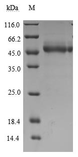
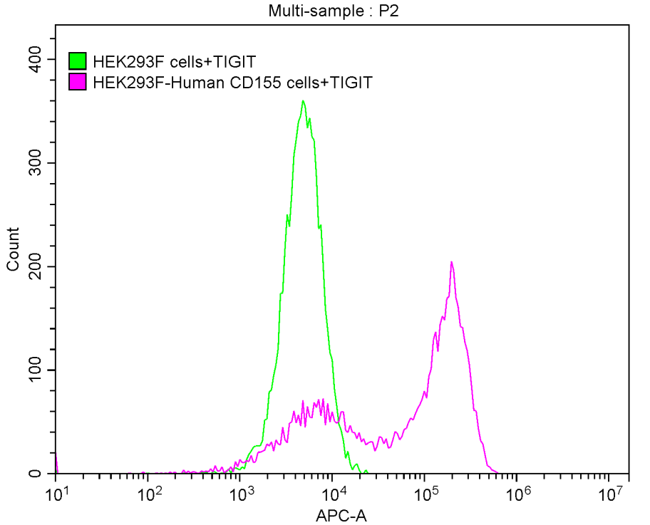
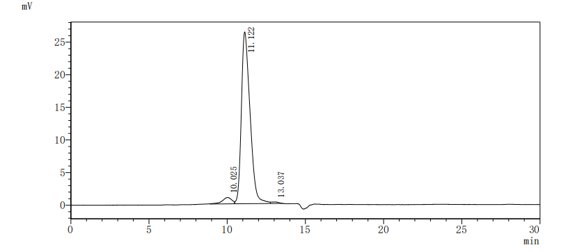
The recombinant human TIGIT protein is expressed in mammalian cells by introducing a plasmid carrying the gene sequence for human TIGIT (22-141aa) and a C-terminal hFc-Myc-tag gene. Its purity is validated to be greater than 95% using SDS-PAGE and SEC-HPLC, and its endotoxin content is measured below 1.0 EU/μg via the LAL assay. This recombinant human TIGIT protein is active, as the FACS assay shows it can bind to 293F cells overexpressing human CD155.
The human TIGIT is a critical immune checkpoint receptor that regulates T cell responses. It is characterized by its structure, which includes an extracellular immunoglobulin variable (IgV) domain, a single transmembrane domain, and a cytoplasmic tail containing two immunoreceptor tyrosine-based inhibitory motifs (ITIMs) [1][2]. This unique configuration allows TIGIT to function as a negative regulator of immune responses, particularly in the context of T cell activation and function.
TIGIT is predominantly expressed on various immune cell types, including effector T cells, memory T cells, Tregs, and NK cells [3][4][5]. Its expression is often associated with T cell exhaustion, a state where T cells lose their ability to proliferate and produce cytokines effectively, particularly in chronic infections and cancer [3][6]. The interaction of TIGIT with its ligands, CD112 (PVRL2) and CD155 (PVR), which are expressed on antigen-presenting cells and some tumor cells, leads to the inhibition of T cell activation and cytokine production, thereby contributing to immune evasion mechanisms in tumors [4][7].
Research has shown that TIGIT's inhibitory effects are mediated through its ITIM domains, which recruit phosphatases that dephosphorylate key signaling molecules involved in T cell activation [2][8]. This mechanism underscores the importance of TIGIT in maintaining immune homeostasis and preventing overactive immune responses that could lead to autoimmunity [4][9]. Furthermore, the modulation of TIGIT expression has been explored as a therapeutic strategy in various diseases, including autoimmune disorders and cancers, where blocking TIGIT can enhance T cell responses and improve therapeutic outcomes [4][10].
References:
[1] G. Chew, T. Fujita, G. Webb, B. Burwitz, H. Wu, J. Reed, et al. Tigit marks exhausted t cells, correlates with disease progression, and serves as a target for immune restoration in hiv and siv infection, Plos Pathogens, vol. 12, no. 1, p. e1005349, 2016. https://doi.org/10.1371/journal.ppat.1005349
[2] L. Bolm, N. Petruch, S. Sivakumar, N. Annels, & A. Frampton. Gene of the month: t-cell immunoreceptor with immunoglobulin and itim domains (tigit), Journal of Clinical Pathology, vol. 75, no. 4, p. 217-221, 2022. https://doi.org/10.1136/jclinpath-2021-207789
[3] C. Zhang, R. Lin, Z. Li, S. Yang, X. Bi, H. Wang, et al. Immune exhaustion of t cells in alveolar echinococcosis patients and its reversal by blocking checkpoint receptor tigit in a murine model, Hepatology, vol. 71, no. 4, p. 1297-1315, 2020. https://doi.org/10.1002/hep.30896
[4] M. Kojima, K. Suzuki, M. Takeshita, M. Ohyagi, M. Iizuka, H. Yamane, et al. Anti-human-tigit agonistic antibody ameliorates autoimmune diseases by inhibiting tfh and tph cells and enhancing treg cells, Communications Biology, vol. 6, no. 1, 2023. https://doi.org/10.1038/s42003-023-04874-3
[5] D. Ozmadenci, J. Narayanan, J. Andrew, M. Ojalill, A. Barrie, S. Jiang, et al. Tumor fak orchestrates immunosuppression in ovarian cancer via the cd155/tigit axis, Proceedings of the National Academy of Sciences, vol. 119, no. 17, 2022. https://doi.org/10.1073/pnas.2117065119
[6] T. Iwasaki. Clinical significance of the expression of foxp3 and tigit in merkel cell carcinoma, Scientific Reports, vol. 13, no. 1, 2023. https://doi.org/10.1038/s41598-023-40050-7
[7] N. Stanietsky, H. Šimić, J. Arapović, A. Toporik, O. Levy, A. Novik, et al. The interaction of tigit with pvr and pvrl2 inhibits human nk cell cytotoxicity, Proceedings of the National Academy of Sciences, vol. 106, no. 42, p. 17858-17863, 2009. https://doi.org/10.1073/pnas.0903474106
[8] C. Abram and C. Lowell. Convergence of immunoreceptor and integrin signaling, Immunological Reviews, vol. 218, no. 1, p. 29-44, 2007. https://doi.org/10.1111/j.1600-065x.2007.00531.x
[9] Q. Luo, P. Fu, Y. Guo, B. Fu, Y. Guo, Q. Huang, et al. Increased tigit+pd‑1+cxcr5‑cd4+t cells are associated with disease activity in rheumatoid arthritis, Experimental and Therapeutic Medicine, vol. 24, no. 4, 2022. https://doi.org/10.3892/etm.2022.11579
[10] W. Tang, J. Chen, T. Ji, & X. Cong. Tigit, a novel immune checkpoint therapy for melanoma, Cell Death and Disease, vol. 14, no. 7, 2023. https://doi.org/10.1038/s41419-023-05961-3
There are currently no reviews for this product.