Call us
301-363-4651 (Available 9 a.m. to 5 p.m. CST from Monday to Friday)
Cancer Testis Antigens (CTAs) are proteins primarily expressed in testicular tissue but not in other normal tissues. However, CTAs show heightened expression in various tumors, with many having tumorigenic properties. Among them, Cancer testis antigen-83 (CT83), also known as KK-LC-1, stands out. Initially identified in a 2006 lung cancer study, CT83 exhibits differential expression across tumors like gastric, breast, and liver cancers. PubMed data indicate limited reports on CT83, and its role in tumors remains unclear. Despite the scarcity of commercial monoclonal antibodies against CT83, clinical studies have begun, sparking interest among drug research institutions in its potential as a novel tumor research target.
CT83, also known as CXorf61 or KK-LC-1, belongs to the Cancer/Testis Antigens (CTAs) family. Originally identified from a lung cancer patient's cell line, it's often referred to as Kita-Kyushu lung cancer antigen-1 (KK-LC-1). Located on chromosome Xq22, the CT83 gene encodes a 113-amino acid protein. Despite the lack of a clear understanding of its structure, its discovery in 1991 marked a significant advancement in tumor/testis antigen research, with over 200 CTAs antigens identified, such as MAGE-A1, NY-ESO-1/CTAG1A, NY-SAR-35/FMR1NB, CT7, SSX2, SCP1, OY-TES-1, PASD1, SLCO6A1, and KK-LC-1, among others. Ongoing investigation into their immunogenicity continues to expand our understanding of their potential in cancer therapy [1-3].
Cancer-testis antigens (CTAs) are proteins mainly found in male testicular tissue but are significantly upregulated in tumors, linking to tumor onset and spread. Notably, CTAs like CT10, CT45, and CT83 are elevated in various cancers, including lung, liver, and breast cancer (Figure 1) [3]. Despite lacking a full understanding of its role, CT83, a member of the CTA family, is found highly expressed in different tumors. Research suggests increased CT83 levels enhance migration, invasion, proliferation, and epithelial-mesenchymal transition (EMT) capacity in liver cancer cells. Consequently, CT83 is considered as a potential target for tumor immunotherapy [4-6].
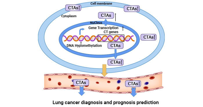
Figure 1. CTAs is closely related to tumor [3]
CT83 is highly expressed in many tumors due to promoter demethylation and activation by the oncogenic transcription factor STAT3. However, the specific factors activating STAT3 and promoting CT83 expression require further investigation. Nevertheless, CT83 overexpression significantly influences cancer cell migration and invasion, enhancing malignant behaviors. Gene knockout studies confirm its essential regulatory role in these processes. Ongoing research aims to uncover the intricate signaling mechanisms underlying CT83's involvement in tumorigenesis [7-9].
A recent study found that CT83 overexpression boosts the activity of cancer-related proteins like ERK, HIF-1a, HIF-2a, and VE-Cadherin in cervical cancer cells. When CT83 expression is inhibited, the levels of these proteins decrease (Figure 2) [6, 10-11, 20]. The ERK pathway, vital for cell growth and cancer development, is closely linked to tumor formation and spread. Transcription factors HIF-1a and HIF-2a activate genes associated with tumor cell movement, attachment, and blood vessel growth. VE-Cadherin helps tumor cells stick together and form new blood vessels [11]. Additionally, studies on CT83 in other cancers suggest it may promote tumor growth by interacting with certain genes like HSPB8 and CDH5 [18-20]. In summary, CT83 appears to influence several key pathways in cancer development, potentially driving tumor progression by altering gene activity and signaling pathways.
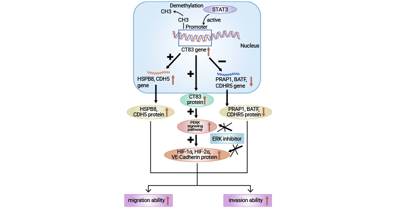
Figure 2. CT83-related signaling pathway [20]
CT83, a tumor-specific antigen, has received limited attention in research both domestically and internationally. However, existing literature suggests its potential significance as a target for future tumor studies. Studies indicate abnormal expression of CT83 in various cancers like lung, gastric, breast, and liver cancer, closely linked to tumor onset and progression. Scholars are increasingly interested in CT83's role and mechanisms in tumors, making it a growing focus of research attention.
Analysis of relevant literature reveals that CT83 activation correlates with reduced survival rates in patients with triple negative breast cancer (TNBC), as shown by bioinformatics analysis. This aberrant activation disrupts 16 crucial cell cycle regulatory genes, including CCNB2, CDC20, CDC25A, CDC25B, CDK1, CHEK1, ESPL1, MAD2L1, MCM2, MCM3, MCM5, MCM6, MYC, ORC6, PLK1, and PTTG1, forming a signaling network driving TNBC progression [21]. CDKs (e.g., CDK1, CDK2, CDK4, CDK6), CDC25 phosphatase, and the oncogenic transcription factor MYC play pivotal roles in cell cycle regulation. Additionally, studies suggest that KK-LC-1, produced via the MAL2/MUC1-C/PI3K/AKT/mTOR pathway, modulates TNBC cell behavior (Figure 3) [22]. Consequently, CT83's synergistic activation of these oncogenic cell cycle regulatory genes likely contributes to TNBC initiation and progression.
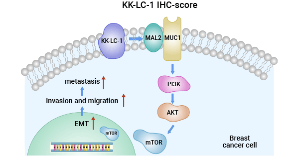
Figure 3. CT83/KK-LC-1 regulates TNBC via MAL2/MUC1-C/PI3K/AKT/mTOR Pathway [22]
Research indicates that CT83/KK-LC-1-specific TCRαβ-CD8 γδT cells effectively target lung cancer cells like F1121L, enhancing anticancer responses via IFN-γ secretion. Moreover, CT83 expression in lung cancer is linked to poor prognosis genes (CPS1, RHOV, TNNT1, FAM83A, JGF2BP1, and GRIN2A) and immune checkpoint molecules (CTLA4, PD1/CD274, IDO1, TDO2, LAG3, and TIGIT) associated with high lung adenocarcinoma risk. Suppressing CT83 in lung cancer cells may improve the immune microenvironment, aiding immune system clearance of lung cancer and boosting patient prognosis. However, CT83's precise role in lung cancer warrants further investigation [23].
Research indicates a high expression of CT83 in Helicobacter pylori-positive gastric cancer patients, suggesting a strong association between them. Elevated titers of Helicobacter pylori IgG in CT83-expressing gastric cancer patients further support CT83 as a potential therapeutic target. Gene expression profiling reveals involvement of key mRNAs and proteins, including ADGRG7, CT83, and MMP12, in various biological processes crucial for early gastric cancer progression. Survival analysis models based on these factors show promising predictive performance, offering valuable insights for clinical decision-making. Therefore, CT83 plays a crucial role in elucidating the interaction between gastric cancer and Helicobacter pylori, guiding targeted therapies, and evaluating prognosis [24].
CT83 is markedly upregulated in hepatocellular carcinoma tissues, affecting the Presenilin-1/Notch1/Hes1 pathway. This pathway regulates liver cancer cell proliferation, migration, invasion, and epithelial-mesenchymal transition (EMT). Notch signaling, crucial for tissue development, drives liver cancer progression. Notch1, predominant in hepatocytes, correlates with liver cancer aggressiveness and poor prognosis. Additionally, CT83 activation via CpG island hypomethylation is linked to liver cancer metastasis. Thus, CT83 holds potential as a biomarker for liver cancer diagnosis and prognosis [25].
In addition, CT83 is overexpressed in various cancers, including melanoma, colon cancer, cervical cancer, esophageal cancer, and pancreatic cancer, garnering considerable attention in medical research [26-29]. Studies reveal a link between CT83's high expression and specific molecular mechanisms, particularly alterations in DNA methylation within its promoter region, often concurrent with STAT3 activation. This suggests that CT83's elevated expression may result from common epigenetic regulation involving promoter demethylation. While CT83 holds potential as a cancer diagnostic marker, prognostic indicator, and immunotherapy target, further validation through rigorous investigation is necessary.
Currently, three anti-cancer drugs targeting CT83/KKLC1 are in clinical trials for breast cancer, lung cancer, and gastric cancer. They work by enhancing immune responses. Major contributors include the National Cancer Institute, T-cure Bioscience, Inc., and Shanghai Jingshan Biotechnology Co., Ltd. At the 2024 AACR meeting, Company CDR-Life Inc. from Swiss presented encouraging preclinical data on TCE antibodies against KK-LC-1/HLA-A01, showing strong anti-tumor effects. Despite challenges with non-target peptides, the TCE antibody effectively targets cancer cells while sparing healthy ones. In vitro studies confirm its specificity and activation potential for the target peptide [30]. These findings support CT83-targeted therapy and offer hope for diverse patients, indicating a promising direction for future cancer treatment research.
CT83/KK-LC-1, a member of the Cancer/Testis Antigens (CTAs) family, is implicated as a promising therapeutic target across various cancers, including breast, lung, and gastric cancers. Its aberrant expression in tumors suggests a role in tumorigenesis and progression. Clinical trials targeting CT83/KK-LC-1 are underway, highlighting its potential in cancer therapy. Ongoing research aims to elucidate its precise mechanisms and evaluate its efficacy as a diagnostic marker and immunotherapy target, reflecting its growing significance in cancer research and treatment.
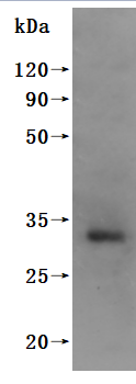
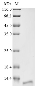
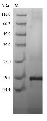
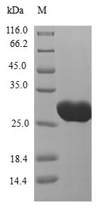
In order to assist pharmaceutical companies in the clinical research of CT83 in various tumors, such as breast cancer, lung cancer, and gastric cancer, etc., CUSABIO is launching the CT83 protein product to help you explore the mechanism of CT83 or its potential clinical value.
CUSABIO protein CT83
References
[1] Bai, Rui, and Cheng Yuan. "Kita-kyushu lung cancer antigen-1 (KK-LC-1): a promising cancer testis antigen." Aging and Disease 13.4 (2022): 1267.
[2] Hsu, Robert, et al. "Molecular characterization of Kita-Kyushu lung cancer antigen (KK-LC-1) expressing carcinomas." Oncotarget 12.25 (2021): 2449.
[3] Yang, Ping, et al. "Cancer/testis antigens as biomarker and target for the diagnosis, prognosis, and therapy of lung cancer." Frontiers in Oncology 12 (2022): 864159.
[4] Zhou, Xingchun, et al. "Heterogeneous expression of CT10, CT45 and GAGE7 antigens and their prognostic significance in human breast carcinoma." Japanese Journal of Clinical Oncology 43.3 (2013): 243-250.
[5] Chen, Zhiqiang, et al. "Hypomethylation‐mediated activation of cancer/testis antigen KK‐LC‐1 facilitates hepatocellular carcinoma progression through activating the Notch1/Hes1 signalling." Cell Proliferation 52.3 (2019): e12581.
[6] Norberg, Scott, et al. "A phase I trial of T-cell receptor gene therapy targeting KK-LC-1 for gastric, breast, cervical, lung and other KK-LC-1 positive epithelial cancers." (2022): TPS2678-TPS2678.
[7] Kang, Yanli, et al. "Cancer-testis antigen KK-LC-1 is a potential biomarker associated with immune cell infiltration in lung adenocarcinoma." BMC cancer 22.1 (2022): 834.
[8] Chen, Zhiqiang, et al. "Hypomethylation‐mediated activation of cancer/testis antigen KK‐LC‐1 facilitates hepatocellular carcinoma progression through activating the Notch1/Hes1 signalling." Cell Proliferation 52.3 (2019): e12581.
[9] Mabjeesh, Nicola J., and S. Amir. "Hypoxia-inducible factor (HIF) in human tumorigenesis." Histology and histopathology (2007).
[10] Marcinkowski, Bridget, et al. "Preclinical characterization of a KK-LC-1-specific T cell receptor for the treatment of epithelial cancers." Cancer Research 79.13_Supplement (2019): 1429-1429.
[11] Kim, Min Kyu, et al. "Clinical significance of HIF-2α immunostaining area in radioresistant cervical cancer." Journal of Gynecologic Oncology 22.1 (2011): 44.
[12] Gul, Samina, et al. "Stemness signature and targeted therapeutic drugs identification for Triple Negative Breast Cancer." Scientific Data 10.1 (2023): 815.
[13] Zeng, Yaoying, et al. "Integrating Network Pharmacology, Molecular Docking, and Experimental Validation to Investigate the Mechanism of (−)-Guaiol Against Lung Adenocarcinoma." Medical Science Monitor: International Medical Journal of Experimental and Clinical Research 28 (2022): e937131-1.
[14] Chen, Chen, et al. "Multiomics analysis reveals CT83 is the most specific gene for triple negative breast cancer and its hypomethylation is oncogenic in breast cancer." Scientific reports 11.1 (2021): 12172.
[15] Gao, Junyi, et al. "Up‐regulation of CDHR5 expression promotes malignant phenotype of pancreatic ductal adenocarcinoma." Journal of Cellular and Molecular Medicine 24.21 (2020): 12726-12735.
[16] Inoue-Shibui, Aya, et al. "A novel deletion in the C-terminal region of HSPB8 in a family with rimmed vacuolar myopathy." Journal of Human Genetics 66.10 (2021): 965-972.
[17] Li, Qingyang, et al. "Natural high-avidity T-cell receptor efficiently mediates regression of cancer/testis antigen 83 positive common solid cancers." Journal for Immunotherapy of Cancer 10.7 (2022).
[18] Marcinkowski, Bridget, et al. "Cancer targeting by TCR gene-engineered T cells directed against Kita-Kyushu Lung Cancer Antigen-1." Journal for immunotherapy of cancer 7 (2019): 1-9.
[19] Lin, Min, et al. "Recent advances on the molecular mechanism of cervical carcinogenesis based on systems biology technologies." Computational and Structural Biotechnology Journal 17 (2019): 241-250.
[20] Qiao Yingnan. The mechanisms underlying regulation of proto-oncogene CT83 expression and promotion of cervical cancer cell migration and invasion [D]. Soochow University, 2022.
[21] Chen, Chen, et al. "Multiomics analysis reveals CT83 is the most specific gene for triple negative breast cancer and its hypomethylation is oncogenic in breast cancer." Scientific reports 11.1 (2021): 12172.
[22] Zhu, Xudong, et al. "Targeting KK-LC-1 inhibits malignant biological behaviors of triple-negative breast cancer." Journal of Translational Medicine 21.1 (2023): 184.
[23] Ichiki, Yoshinobu, et al. "Development of adoptive immunotherapy with KK‐LC‐1‐specific TCR‐transduced γδT cells against lung cancer cells." Cancer Science 111.11 (2020): 4021-4030.
[24] Hu, Yeting, et al. "Quantitative Analysis on Molecular Characteristics Evolution of Gastric Cancer Progression and Prognosis." Advanced Biology 7.10 (2023): 2300129.
[25] Chen, Zhiqiang, et al. "Hypomethylation‐mediated activation of cancer/testis antigen KK‐LC‐1 facilitates hepatocellular carcinoma progression through activating the Notch1/Hes1 signalling." Cell Proliferation 52.3 (2019): e12581.
[26] Otsuka, Toshikazu, et al. "Detection of Kita-Kyushu Lung Cancer Antigen-1, a Cancer/Testis Antigen, in the Stomach Close to a Cancerous Condition." Journal of Cancer 13.14 (2022): 3526.
[27] Fukuyama, Takashi, et al. "Expression of KK-LC-1, a cancer/testis antigen, at non-tumour sites of the stomach carrying a tumour." Scientific reports 8.1 (2018): 6131.
[28] Bai, Rui, and Cheng Yuan. "Kita-kyushu lung cancer antigen-1 (KK-LC-1): a promising cancer testis antigen." Aging and Disease 13.4 (2022): 1267.
[29] Marcinkowski, Bridget, et al. "Cancer targeting by TCR gene-engineered T cells directed against Kita-Kyushu Lung Cancer Antigen-1." Journal for immunotherapy of cancer 7 (2019): 1-9.
[30] Scheifele, Fabian, et al. "Abstract LB442: Novel antibodies against a KK-LC-1-derived peptide presented on HLA-A* 01 on tumor cells." Cancer Research 84.7_Supplement (2024): LB442-LB442.


Comments
Leave a Comment