Call us
301-363-4651 (Available 9 a.m. to 5 p.m. CST from Monday to Friday)
CASP8, full name caspase-8. It is a key mediator of programmed cell death, eliminating damaged abnormal cells and maintaining normal cell renewal and immune homeostasis in human body.
Inactivated CASP8 induces tumors and autoimmune diseases, while overactivation causes inflammatory and neural damage. CASP8 modulators are under preclinical and early clinical development for cancer and inflammatory disorders.
Recombinant Human Caspase-8 (CASP8), partial (CSB-EP621791HUc7)
Validated Data

(Tris-Glycine gel) Discontinuous SDS-PAGE (reduced) with 5% enrichment gel and 15% separation gel.
CASP8 Antibody (CSB-PA18109A0Rb)
Validated Data
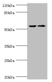
Western blot
All lanes: CASP8 antibody at 2µg/ml
Lane 1: ACCM whole cell lysate
Lane 2: LO2 whole cell lysate
Secondary
Goat polyclonal to rabbit IgG at 1/10000 dilution
Predicted band size: 56, 54, 46, 58, 28, 26, 33, 31, 62 kDa
Observed band size: 56 kDa
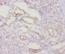
Immunohistochemistry of paraffin-embedded human kidney tissue using CSB-PA18109A0Rb at dilution of 1:20
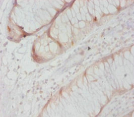
Immunohistochemistry of paraffin-embedded human colon cancer using CSB-PA18109A0Rb at dilution of 1:20
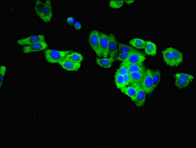
Immunofluorescent analysis of HepG2 cells using CSB-PA18109A0Rb at dilution of 1:100 and Alexa Fluor 488-congugated AffiniPure Goat Anti-Rabbit IgG(H+L)
Human Caspase 8(Casp-8) ELISA Kit (CSB-E13636h)
Validated Data
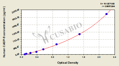
Code: CSB-E13636h
Size: 96T,5×96T,10×96T
Sensitivity: 7.81 pg/ml
Detection Range: 31.25 pg/mL-2000 pg/mL
These standard curves are provided for demonstration only. A standard curve should be generated for each set of samples assayed.
The following CASP8 reagents supplied by CUSABIO are manufactured under a strict quality control system. Multiple applications have been validated and solid technical support is offered.
CASP8 Antibodies for Homo sapiens (Human)
| Code | Product Name | Species Reactivity | Application |
|---|---|---|---|
| CSB-PA18109B0Rb | CASP8 Antibody, HRP conjugated | Human | ELISA |
| CSB-PA004553GA01HU | CASP8 Antibody | Human | ELISA,WB,IHC |
| CSB-PA18109A0Rb | CASP8 Antibody | Human | ELISA, WB, IHC, IF |
| CSB-PA982156 | CASP8 Antibody | Human | ELISA,WB |
| CSB-PA000009 | Cleaved-CASP8 (D384) Antibody | Human | WB, ELISA |
| CSB-PA000653 | Phospho-CASP8 (Y380) Antibody | Human | WB, ELISA |
| CSB-PA001234 | CASP8 Antibody | Human,Mouse,Rat | WB, IHC, ELISA |
| CSB-PA060299 | Phospho-CASP8 (S347) Antibody | Human,Rat | WB, IHC, ELISA |
| CSB-MA080171 | CASP8 Monoclonal Antibody | Human,Mouse,Rat | ELISA,WB,IHC |
| CSB-PA080234 | CASP8 Antibody | Human,Mouse,Rat | WB, IHC |
| CSB-PA181094ZA01HU | CASP8 Antibody | Homo sapiens | ELISA, WB (ensure identification of antigen) |
| CSB-RA549188A0HU | CASP8 Recombinant Monoclonal Antibody | Human | ELISA, WB, IHC, IF, FC |
CASP8 Proteins for Mus musculus (Mouse)
| Code | Product Name | Source |
|---|---|---|
| CSB-YP004553MO CSB-EP004553MO CSB-BP004553MO CSB-MP004553MO CSB-EP004553MO-B |
Recombinant Mouse Caspase-8 (Casp8) | Yeast E.coli Baculovirus Mammalian cell In Vivo Biotinylation in E.coli |
CASP8 Proteins for Homo sapiens (Human)
| Code | Product Name | Source |
|---|---|---|
| CSB-BP621791HU CSB-MP621791HU CSB-EP621791HU-B |
Recombinant Human Caspase-8 (CASP8), partial | Baculovirus Mammalian cell In Vivo Biotinylation in E.coli |
| CSB-EP621791HU | Recombinant Human Caspase-8 (CASP8), partial | E.coli |
| CSB-EP621791HUc7 | Recombinant Human Caspase-8 (CASP8), partial | E.coli |
| CSB-YP621791HU | Recombinant Human Caspase-8 (CASP8), partial | Yeast |
CASP8 ELISA Kit for Homo sapiens (Human)
| Code | Product Name | Sample Types | Sensitivity |
|---|---|---|---|
| CSB-E13636h | Human Caspase 8(Casp-8) ELISA Kit | serum, plasma, cell lysates | 7.81 pg/ml |

