CEACAM5, a prominent tumor marker on various solid tumors, was highlighted at the European Lung Cancer Congress 2023 (ELCC2023). The event revealed encouraging outcomes of CEACAM5-targeted drug Tusamitamab ravtansine (SAR408701) in treating CEACAM5-positive advanced non-squamous NSCLC. SAR408701, a pioneering ADC, features a humanized monoclonal antibody (IgG1) targeting CEACAM5, along with a cleavable linker and the potent anti-microtubulin drug medenosin (DM4). With its rapid progress, SAR408701 targeting CEACAM5 is expected to be available soon.
Recent research has highlighted CEACAM5 as a promising target for new drugs. Several drugs targeting CEACAM5 are in development, with collaborations with international pharmaceutical companies and research institutions such as Promise Biologics, Roche, Sanofi, and others. Today, let's explore what CEACAM5 is and why it's making waves in cancer research!
4. The Clinical Research of CEACAM5 Targeted Therapy
5. CUSABIO CEACAM5 Recombinant Proteins & Antibodies for Research Use
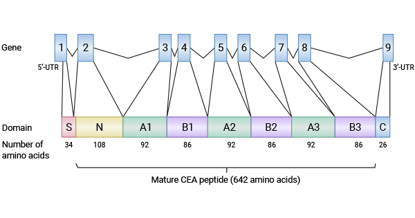
CEACAM5 (carcinoembryonic antigen-related cell adhesion molecule-5), also known as CD66e or CEA, is a part of the CEA family (Click here to see "What is the CEA family?"). CEACAM5 is characterized by seven extracellular Ig chains and a glucosylphosphatidylinositol chain, exhibiting a domain structure of N-A1-B1-A2-B2-A3-B3 (Figure 1) [1]. Among the CEA gene family, CEACAM1, CEACAM5, CEACAM6, and CEACAM7 have crucial roles in tumorigenesis. While CEACAM1 and CEACAM7 are commonly down-regulated or absent in many tumors, indicating their function as tumor suppressor genes, CEACAM5 and CEACAM6 are often elevated in half of the tumors, acting as oncogenes [1-3].
CEACAM5 is widely found on the surfaces of different cell types, including endothelial, epithelial, and immune cells. It primarily functions as a cell adhesion molecule, promoting both homogeneous and heterogeneous adhesion in normal tissues. Similar to CEACAM6, it helps tumor cells evade apoptosis (Check the earlier CEACAM6 article here). Increased CEACAM5 and CEACAM6 levels can disturb cell polarity and hinder the maturation and differentiation of various cells, possibly contributing to tumorigenesis. Currently, CEACAM5 is used as a tumor marker for diagnosing various tumors and predicting their progression, deterioration, and subclinical metastasis [1, 4-5].
Figure 1. Structure of CEACAM5 [5]
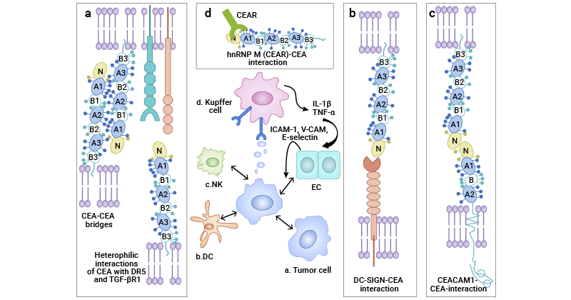
CEACAM5 is highly expressed in most malignant tumors and is linked to tumorigenesis, invasion, and metastasis. Its mechanism of action, as part of CEACAMs, can be summarized in three key aspects: (i). Anti-adhesion - crucial for tumor invasion and metastasis, driving malignant tumor progression; (ii). Extracellular matrix (ECM) degradation - vital for tumor cell migration, with excessive CEACAM expression hindering apoptosis and disrupting cell structure; (iii). Activation of tumor signaling pathways-CEACAMs activate integrin signaling pathways, including elemental ILK and the PI3K. Notably, only GPI-anchored CEACAMs can bind to the α5β1 integrin receptor, exerting biological effects. Soluble CEACAMs lacking the GPI-anchor lose their binding ability, leading to apoptosis by blocking cell survival signaling from extracellular to intracellular processes (Figure 2) [6].
Figure 2. The mechanisms of CEACAM5 in tumors [6]
Platelets play a crucial role in tumor spreading and metastasis. CEACAM5/CEA, catabolized by PI-PLC/D, releases soluble sCEA, activating the c-Src kinase/MAP-ERK kinase pathway. This promotes cell adhesion, proliferation, migration, and microvascularization. Research shows that the extracellular segment of CEACAM5 inhibits certain platelet functions. Notably, MMP-12, MMP-2, MMP-7, and MMP-9 do not affect platelets through cleaving CEACAM5. This finding enhances our understanding of how CEACAM5 impacts platelets and tumor angiogenesis [7].
CEACAM5 is commonly used in early detection, diagnosis, and prognosis of colorectal cancer (CRC). Its elevated levels suggest potential disease progression. Studies connect increased CEACAM5 with higher recurrence risk in CRC patients. Combining the CDX2 test is suggested for comprehensive prognosis [9]. Additionally, positive SRCAP and CEACAM5 expression relate to higher postoperative recurrent metastasis rates, serving as predictors for recurrence after radical CRC surgery [10]. In conclusion, while crucial for early CRC management, CEACAM5 is often combined with other markers for accurate disease assessment.
Studies showed higher CEACAM5 expression in lung adenocarcinoma. Ualcan database analysis revealed significant differences between CEACAM5-positive and negative groups in various clinical factors. Kaplan-Meier survival analysis indicated lower survival rates in CEACAM5-positive patients. COX regression analysis identified CEACAM5, clinical stage, and lymph node metastasis as prognostic risk factors. Additionally, a positive correlation was found between CEACAM5, CEACAM6, and CEACAM1 in lung adenocarcinoma, suggesting their role in cancer development. These findings imply that high CEACAM5 expression is linked to poor prognosis in lung adenocarcinoma and may be a treatment target in lung cancer research [11].
the MGd1 monoclonal antibody targets CEACAM5, allowing the assessment of its expression in gastric cancer patients for prognosis prediction. Analysis of 643 gastric tissues revealed increasing CEACAM5 expression from normal gastric mucosa to gastric cancer, emphasizing its role in cancer development. CEACAM5-positive non-cancerous gastric tissues were more prone to developing gastric cancer, suggesting its potential as a biomarker. Survival analysis of 143 gastric cancer patients indicated a positive link between CEACAM5 expression and deep cancer infiltration, particularly in advanced stages (IIIA-IV). Thus, CEACAM5 serves as an independent prognostic indicator for gastric cancer, correlating with poorer survival [12].
RT-qPCR analysis examined KRT19 mRNA and CEACAM5 mRNA-labeled CTC levels in preoperative peripheral blood of breast cancer patients. Results showed higher CTC KRT19-positive and CTC CEACAM5-positive expression rates in metastatic breast cancer patients (stage IV) compared to earlier stages (0-III). These findings support KRT19 and CEACAM5 as potential biomarkers for predicting breast cancer metastasis. In breast cancer, markers like KRT19 and CEACAM5 in serum or tumor tissue have been used to detect metastasis and predict prognosis [13].
Research on CEACAM5 in various cancers has increased in recent years. In Mucoepidermoid Carcinoma (MEC) of the salivary glands, high CEACAM5 and PCNA expression indicates increased malignancy, aggressiveness, lymph node metastasis, and poor prognosis. Evaluating CEACAM5 and PCNA levels in salivary gland tumor patients can help determine malignancy and prognosis, guiding treatment decisions [14]. Additionally, CEACAM5 is overexpressed in pancreatic, gallbladder, female reproductive system, medullary thyroid carcinoma, bladder, and prostate tumors, highlighting its potential as a versatile tumor marker and immunotherapy target [15-20]!
Currently, there are 15 drugs related to CEACAM 5 in development, including monoclonal antibodies, bispecific antibodies, CAR T cell therapies, trispecific antibodies, diagnostic radiopharmaceuticals, and therapeutic vaccines. Sanofi's Tusamitamab ravtansine, in phase III trials for non-small cell lung cancer, is the most advanced. Other drugs are in various clinical phases, such as the bispecific antibodies: Labetuzumab govitecan (CEACAM 5 x TOP1), BGB-B167 (CEACAM 5 x 4-1BB), M-9140 (CEACAM 5 x TOP1), CT-109 (CEACAM 5 x CEACAM6), YB-200 (CEACAM1 x CEACAM 5), CEACAM5 x CD3; triple-specific antibodies: PM-4008 (CEACAM 5 x CD3 x CEACAM6); CAR T: Anti- CEA CAR T cell therapy; Vaccine: Cancer vaccine (CEACAM 5 x MUC1); diagnostic radiopharmaceuticals: TF 2-targeted cancer therapy, etc. These developments suggest a promising future for CEACAM5 as a tumor target.
CEACAM5, a cell adhesion molecule and member of the CEA family, is overexpressed in various cancers, notably colorectal, lung, gastric, and breast cancers. Its high expression often correlates with aggressive tumor behavior and poor prognosis. Studies suggest its utility as a diagnostic marker for cancer detection and prognostic evaluation. Ongoing drug development targeting CEACAM5 includes monoclonal antibodies, CAR T cell therapies, and bispecific antibodies, indicating its potential as a valuable therapeutic target in cancer research.
To fully support researchers and pharmaceutical companies in their research on CEACAM5 in tumors, CUSABIO presents CEACAM5 active proteins & antibodies to support your research on the mechanism of CEACAM5 or its potential clinical value.
CUSABIO CEACAM5 Protein
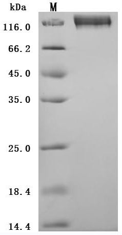
The high purity is greater than 95% as determined by SDS-PAGE.
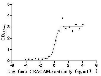
Immobilized Human CEACAM5 at 2μg/mL can bind Anti-CEACAM5 recombinant antibody (CSB-RA005165MA1HU). The EC50 is 0.8955-1.719 ng/mL.
CUSABIO CEACAM5 antibody
CEACAM5/CEACAM6 Recombinant Monoclonal Antibody (Code: CSB-RA005165MA2HU)
References
[1] Zhou, Xile. Near-infrared fluorescent imaging of pancreatic cancer in mice using a novel antibody to CEACAM5. Diss. Dissertation, Kiel, Christian-Albrechts-Universität zu Kiel, 2021, 2021.
[2] Grunnet, M., and J. B. Sorensen. "Carcinoembryonic antigen (CEA) as tumor marker in lung cancer." Lung cancer 76.2 (2012): 138-143.
[3] Hammarström, Sten. "The carcinoembryonic antigen (CEA) family: structures, suggested functions and expression in normal and malignant tissues." Seminars in cancer biology. Vol. 9. No. 2. Academic Press, 1999.
[4] The Atlas of Genetics and Cytogenetics in Oncology and Haematology “https://atlasgeneticsoncology.org/gene/624/alpg-(alkaline-phosphatase-germ-cell)”.
[5] Blumenthal, Rosalyn D., et al. "Expression patterns of CEACAM5 and CEACAM6 in primary and metastatic cancers." BMC cancer 7 (2007): 1-15.
[6] Beauchemin, Nicole, and Azadeh Arabzadeh. "Carcinoembryonic antigen-related cell adhesion molecules (CEACAMs) in cancer progression and metastasis." Cancer and Metastasis Reviews 32 (2013): 643-671.
[7] Wan, Wen, et al. "Platelet Carcinoembryonic Antigen Cell Adhesion Molecule 5 (CEACAM5) as a Possible Novel Diagnostic Tool for Evaluation of Acute Coronary Syndrome." Medical Science Monitor: International Medical Journal of Experimental and Clinical Research 25 (2019): 9864.
[8] Xiao, Yitai, et al. "Identification of a CEACAM5 targeted nanobody for positron emission tomography imaging and near-infrared fluorescence imaging of colorectal cancer." European Journal of Nuclear Medicine and Molecular Imaging (2023): 1-14.
[9] Mohapatra, Shyam S., et al. "Precision medicine for CRC patients in the veteran population: state-of-the-art, challenges and research directions." Digestive diseases and sciences 63 (2018): 1123-1138.
[10] Moon, Seong Won, et al. "Cancer-related SRCAP and TPR mutations in colon cancers." Pathology-Research and Practice 217 (2021): 153292.
[11] Singer, Bernhard B., et al. "Deregulation of the CEACAM expression pattern causes undifferentiated cell growth in human lung adenocarcinoma cells." PloS one 5.1 (2010): e8747.
[12] Zhou, Jinfeng, et al. "Identification of CEACAM5 as a biomarker for prewarning and prognosis in gastric cancer." Journal of Histochemistry & Cytochemistry 63.12 (2015): 922-930.
[13] Wang, Xi-Mei, et al. "KRT19 and CEACAM5 mRNA-marked circulated tumor cells indicate unfavorable prognosis of breast cancer patients." Breast cancer research and treatment 174 (2019): 375-385.
[14] Sai, M. A., and A. N. Feng. "Expression and influence of CEACAM 5 and PCNA in mucinous epidermoid carcinoma of the parotid gland with different clinical pathological features on clinical prognosis." Journal of Hebei Medical University 41.4 (2020): 440.
[15] Govindan, Serengulam V., et al. "CEACAM5-targeted therapy of human colonic and pancreatic cancer xenografts with potent labetuzumab-SN-38 immunoconjugates." Clinical Cancer Research 15.19 (2009): 6052-6061.
[16] Zhang, Xinwen, et al. "CEACAM5 stimulates the progression of non-small-cell lung cancer by promoting cell proliferation and migration." Journal of International Medical Research 48.9 (2020): 0300060520959478.
[17] Baek, Du-San, et al. "A highly-specific fully-human antibody and CAR-T cells targeting CD66e/CEACAM5 are cytotoxic for CD66e-expressing cancer cells in vitro and in vivo." Cancer Letters 525 (2022): 97-107.
[18] Igami, Ko, et al. "Extracellular vesicles expressing CEACAM proteins in the urine of bladder cancer patients." Cancer Science 113.9 (2022): 3120-3133.
[19] Yan, Jie-ke, et al. "CEACAM5 is correlated with Angio/Lymphangiogenesis of Prostatic Lesions." Central European Journal of Medicine 9 (2014): 264-271.
[20] DeLucia, Diana C., et al. "Regulation of CEACAM5 and Therapeutic Efficacy of an Anti-CEACAM5–SN38 Antibody–drug Conjugate in Neuroendocrine Prostate Cancer." Clinical Cancer Research 27.3 (2021): 759-774.
Comments
Leave a Comment