Call us
301-363-4651 (Available 9 a.m. to 5 p.m. CST from Monday to Friday)
A recent article published in the American Journal of Respiratory and Critical Care Medicine reveals that CEACAM6 is a key inhibitor of HO-1 inhibitor that contributes to the development of lung disease [1]. Carcinoembryonic antigen cell adhesion molecules (CEACAMs) belong to the Carcinoembryonic Antigen (CEA) gene family. CEACAMs are a well-studied class of tumor-associated antigens. CEACAMs have been implicated in many cellular processes such as cell adhesion, cell proliferation, angiogenesis, and tumorigenesis.
At this year's AACR annual meeting, Sanofi and LaNova Medicines announced progress in developing CEACAM5 ADC and dual-antibody therapies. Intriguingly, CEACAM6, as a member of the CEA family, researchers discovered a significant link between CEACAM6 and tumor development, emphasizing its importance as a valuable biomarker and a potential therapeutic target in multiple cancers!
1. What is the Carcinoembryonic Antigen (CEA) Gene Family?
3. How's the Mechanism of Action of CEACAM6 in Tumors?
The Carcinoembryonic Antigen (CEA) Gene Family is classified into two primary groups: CEA-related Cell Adhesion Molecule (CEACAM) and Pregnancy Specific-Glycoprotein (PSG). The PSG family comprises PSG1-11, while the CEACAM family includes at least 12 members such as CEACAM1, CEACAM3, CEACAM4, CEACAM5, CEACAM6, CEACAM7, CEACAM8, etc. The CEACAM family has garnered significant attention in cancer research as a tumor-associated antigen. For example, CEACAM1, CEACAM5, and CEACAM7 serve as important targets for drug development. In addition, CEACAM6's high expression in tumors also contributes to its oncogenic potential [1-3].
Carcinoembryonic antigen-related cell adhesion molecule 6 (CEACAM6), also known as CD66c and NCA-90, is a protein belonging to the CEA family of cell adhesion molecules. It is located on chromosome 19 q13.2 and consists of 344 amino acid residues. CEACAMs have an N-terminal immunoglobulin variable domain (IgV), varying number of immunoglobulin constant domains (IgC2), and are anchored to the plasma membrane through either a transmembrane and cytoplasmic domain or a glycophosphatidylinositol (GPI) moiety. They interact with each other through homo-and/or hetero-affinity interactions (eg. CEACAM6-CEACAM8 and CEACAM5-CEACAM6) (Figure 1). CEACAMs also serves as a partner to various receptors including T cells, NK cells, TLR-2, TLR-4, VEGFR1, VEGFR2, VEGFR3, EGFR, insulin receptor, GM-CSFR, and others [2-4].
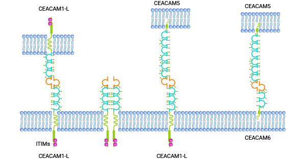
Figure 1. CEACAMs structure and interactions [3]
CEACAM6 is minimally expressed in normal epithelial and vascular endothelial cells, such as granulocytes, T-cells, NK cells. However, its expression is significantly higher in various malignant tumors affecting the colon, stomach, pancreas, breast, female reproductive system, and lung. Extensive research has shown that CEACAM6 contributes to tumor development and progression by promoting cell growth, migration, and invasion, while also inhibiting cell death [5-9].
It also plays a role in facilitating blood vessel formation and inducing drug resistance. Among all carcinoembryonic antigen genes, CEACAM6 stands out as the most notable biological marker for aggressive tumors. Hence, targeting CEACAM6 represents a promising novel treatment strategy to combat tumor metastasis (Figure 2) [5-9].
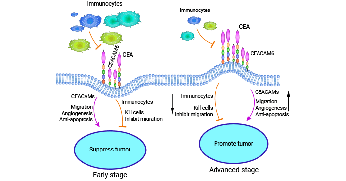
Figure 2. CEACAM6 is a distinctive biological marker in numerous aggressive tumors [5]
Researchers have found that CEACAM6 is associated with altered cell differentiation and organization, tumor invasion and metastasis, angiogenesis, apoptosis inhibition, and tumor suppression. It has been shown to play a key role in regulating the immune response of CD8+ T cells to tumors through the PI3K/Akt signaling pathway. However, further investigation is needed to fully understand the upstream regulators of CEACAM6 in tumor development [10].
Studies have indicated that overexpression of CEACAM6 has implications in the reorganization of the Extracellular Matrix (ECM) and activation of the tumor microenvironment (TME). CEACAM6 up-regulation triggers Src activity, leading to autocrine and insulin-like growth factor IGF-1R activation. Consequently, the PI3K/Akt pathway is regulated, promoting IGF-I paracrine stimulation. The elevated IGF-I expression activates matrix metalloproteinase 2 (MMP-2), which alters the ECM and fosters the formation of a malignant TME [10].
After CEACAM6 aggregates on the cell surface, it associates with Cav1 and enhances Src activity, increasing cellular resistance to apoptosis. In the TME, TGF-β binding to TGFBR2 leads to the activation of TGFBR1, phosphorylation of SMAD3, and formation of a SMAD3-SMAD4 complex. This complex migrates to the nucleus, where it activates the expression of target genes, including CEACAM6. Furthermore, TGF-β has the ability to activate signaling pathways other than AKT/PI3K (Figure 3) [10].
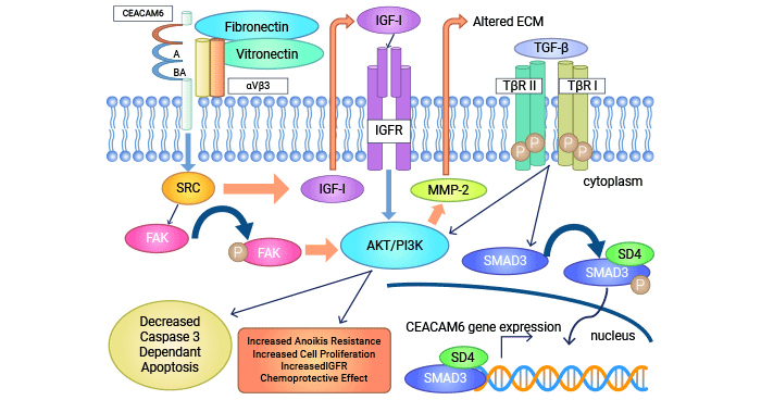
Figure 3. The potential regulation mechanism of CEACAM6 in tumor [10]
Cholangiocarcinoma is a liver-associated malignant tumor arising from bile duct epithelial cells. In patients with intrahepatic cholangiocarcinoma, it suggested that CEACAM6 over-expression was linked to lymph node metastasis, advanced tumor stage, and shorter survival. In TFK-1 cell line, the higher CEACAM6 expression was observed compared to Hucc-T1 and MEC cell lines, indicating increased resistance to Gemcitabine chemotherapy [13-14, 17].
Besides, up-regulated CEACAM6 enhanced tumor growth, invasiveness, and Gemcitabine resistance, while silencing CEACAM6 increased sensitivity to Gemcitabine treatment. These findings highlight CEACAM6's role in evaluating and treating aggressive tumors, making it a potential drug target in clinical applications [13-14, 17].
In pancreatic ductal adenocarcinoma (PDA), over-expression of CEACAM6 contributes to tumor cells evading apoptosis (Anoikis resistance). Anoikis resistance allows cancer cells to survive and spread by evading cell death triggered when they detach from the surrounding tissue. However, silencing CEACAM6 using siRNA reversed anoikis resistance in PDA [15-18].
Dow-regulation of CEACAM6 via siRNA increased cellular sensitivity to cysteine asparaginase-induced tumor-cell death and reduced AKT phosphorylation. In an in situ transplantation model using nude mice, inhibition of CEACAM6 decreased the metastatic potential of pancreatic cancer cell lines. CEACAM6 in PDA may confer resistance to Gemcitabine treatment, which in turn diminishes the effectiveness of therapy [15-18].
CEACAM6 exhibits high expression in breast cancer cells and signifies role in promoting CD34+ angiogenesis. Its expression is correlated with disease progression and resistance to Tamoxifen treatment. Moreover, in HER2+ breast cancer, low CEACAM6 expression suggests sensitivity to Trastuzumab, while CEACAM6 overexpression indicates resistance [19-20]. Consequently, the assessment of CEACAM6 expression levels facilitates the identification of breast cancer patients at elevated risk and enables the customization of treatment strategies to breast cancer.
The expression of CEACAM6 gradually increases from normal tissue to colon adenoma and further to colon cancer. Immunohistochemistry analysis has demonstrated that CEACAM6 can induce cell migration and enhance invasiveness in tumor cells. Particularly in colon cancer, the expression of CEACAM6 and FOXP3 is associated with the density of infiltrating CD3+, CD4+, CD8+, and CD45RO+ T cells. Specifically, CEACAM6 expression is significantly higher in stage III and IV colon cancers compared to stage I and II. The high levels of CEACAM6 and FOXP3 have an inhibitory effect on the infiltration of cytotoxic and memory T cells in colon cancer tissues [6, 21-22].
CEACAM6 was able to increase the migration and invasive ability of gastric cancer cells in vitro, and this alteration was largely inhibited by its monoclonal antibody. In addition, CEACAM6 was able to inhibit apoptosis of gastric cancer cells. In nude mice, overexpression of CEACAM6 increased C-SRC protein activity and formed obvious metastatic foci in liver, lung and other organs compared with negative control group, suggesting that CEACAM6 can participate in gastric cancer development by increasing the expression of proto-oncogene C-SRC [2, 23-24].
CEACAM6 also plays a crucial role in various other cancers, including lung cancer [25], thyroid cancer [26], renal cell carcinoma [27], B-lymphoblastic leukemia [28-29], and multiple myeloma [30-31]. In myeloma cells, blocking CEACAM6 expression using monoclonal antibodies or RNA interference can restore T-cell responses against tumor cells [31].
In B-cell acute lymphoblastic lymphoma, CEACAM6 enhances anoikis resistance by increasing caspase activity and activating the Akt cell survival pathway [29]. In lung cancer cells, the upregulation of CEACAM6 promotes epithelial-mesenchymal transition (EMT) via by inhibitor of growth 5 (ING5) [32]. In renal cell carcinoma, CEACAM6 may exert its effects through the ERK/AKT pathway, leading to increased expression of C-MYC, Survivin, and MMP-9 [27].
Pharmsnap data shows there are several clinical trial drugs targeting CEACAM6 for pancreatic, breast, lung, and colorectal cancers (Table 1). Notably, Immunomedics' Sulesomab has received marketing approval. Previous studies have demonstrated the potential of targeting CEACAM6 in tumor treatment. For example, antibody-drug conjugates against CEACAM6 have been effective in mouse models of pancreatic ductal adenocarcinoma (PDA) [33].
Additionally, a humanized monoclonal antibody's fragment has induced apoptosis in PDA cells by specifically targeting CEACAM6 sites, allowing for lower doses in combination with conventional chemotherapy while maintaining efficacy [34-35]. Overall, as researchers delve deeper into the CEA family, the idea of targeting CEACAM6 might open up new possibilities in the tumors treatment.
| Drug name | Target | Mechanism of action | Indications | Drug status (global) | Type of drug | Institutions |
|---|---|---|---|---|---|---|
| Sulesomab | CEACAM6 | CEACAM6 inhibitor | osteomyelitis | Approved for listing |
Radiolabeled antibodies; Diagnostic radiopharmaceuticals; Monoclonal antibodies |
Immunomedics, Inc. |
| L-DOS-47 | CEACAM6 | CEACAM6 inhibitor |
Pancreatic cancer; Lung cancer; Non-small cell lung cancer |
Clinical Phase 2 | fusion protein | Helix BioPharma Corp. |
| NEO-201 | CEACAM5 + CEACAM6 | CEACAM5 antagonist, CEACAM6 inhibitor |
Squamous cell carcinoma of the head and neck; Non-small cell lung cancer; Cervical cancer; Lung adenocarcinoma; Breast cancer; Colorectal cancer; Pancreatic cancer. |
Clinical phase 1/2 | monoclonal antibody | Precision Biologics, Inc. |
| EBC-129 | CEACAM6 | CEACAM6 inhibitor | solid tumor | Clinical Phase 1 | ADC | Singapore Center for Experimental Drug Development |
| PM-4008 | CD3 + CEACAM6 | CEACAM6 inhibitor | neoplasms | preclinical | trispecific antibody | Biotheus Inc. |
| DNP-002 | CEACAM6 | CEACAM6 inhibitor | solid tumor | preclinical | small molecule chemotherapy | DiNonA, Inc. |
| BAY-1834942 (Deutsches Krebsforschungszentrum, Dkfz) | CEACAM6 | CEACAM6 inhibitor | neoplasms | preclinical | monoclonal antibody | Bayer AG; German Cancer Research Center (Deutsches Krebsforschungszentrum, DKFZ) |
| ICT-109 | CEACAM5 + CEACAM6 | CEACAM5 antagonist, CEACAM6 inhibitor | / | drug discovery | monoclonal antibody | / |
Table 1. CEACAM6 clinical trials for drug development
To fully support researchers and pharmaceutical companies in their research on CEACAM6 in cancers, CUSABIO presents CEACAM6 active protein to support your research on the mechanism of CEACAM6 or its potential clinical value (click for the full list of CEACAM6 products: CEACAM6 proteins; CEACAM6 antibodies; CEACAM6 kits).
CEACAM6 protein
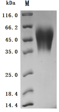
The purity was greater than 95% as determined by SDS-PAGE. (Tris-Glycine gel) Discontinuous SDS-PAGE (reduced) with 5% enrichment gel and 15% separation gel.
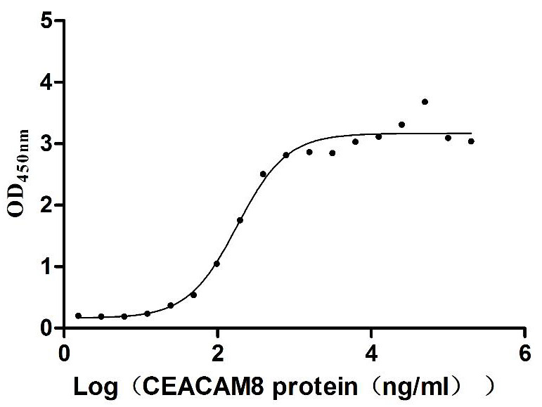
Immobilized Human CEACAM6 at 2 μg/mL can bind Human CEACAM8 (CSB-MP005168HU), the EC50 is 144.7-223.8 ng/mL.
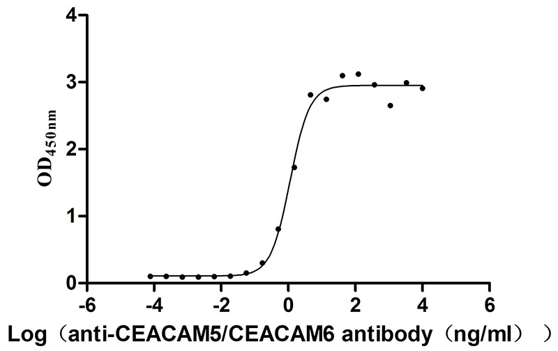
Immobilized Human CEACAM6 at 2 μg/mL can bind Anti-CEACAM5/CEACAM6 recombinant antibody (CSB-RA005165MA2HU), the EC50 is 0.9430-1.377 ng/mL.
CEACAM5/CEACAM6 Recombinant Antibodies
CEACAM5/CEACAM6 Recombinant Monoclonal Antibody (Code: CSB-RA005165MA2HU)
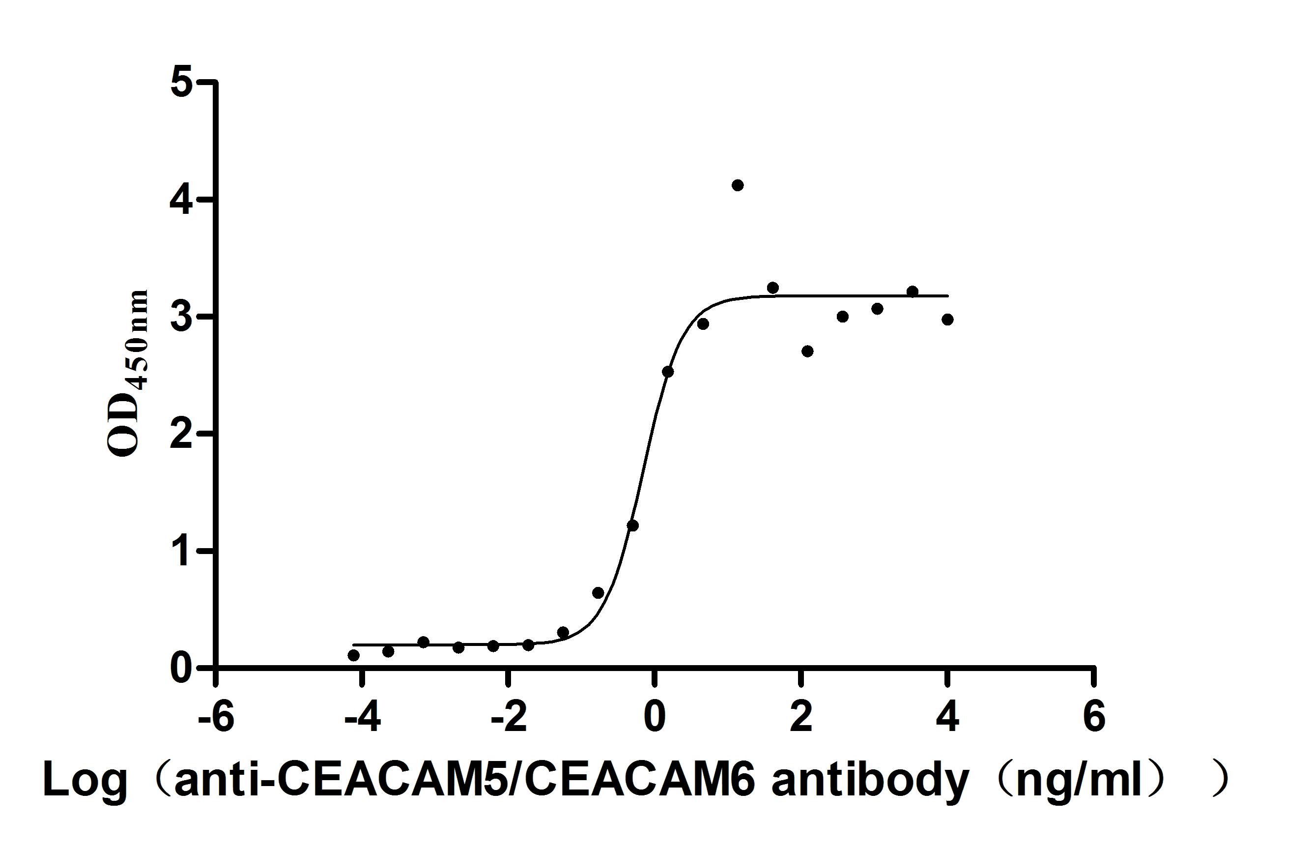
Measured by its binding ability in a functional ELISA. Immobilized Human CEACAM5 at 2μg/mL can bind Anti-CEACAM5/CEACAM6 recombinant antibody (CSB-RA005165MA2HU), the EC50 is 0.4282-1.151 ng/mL.
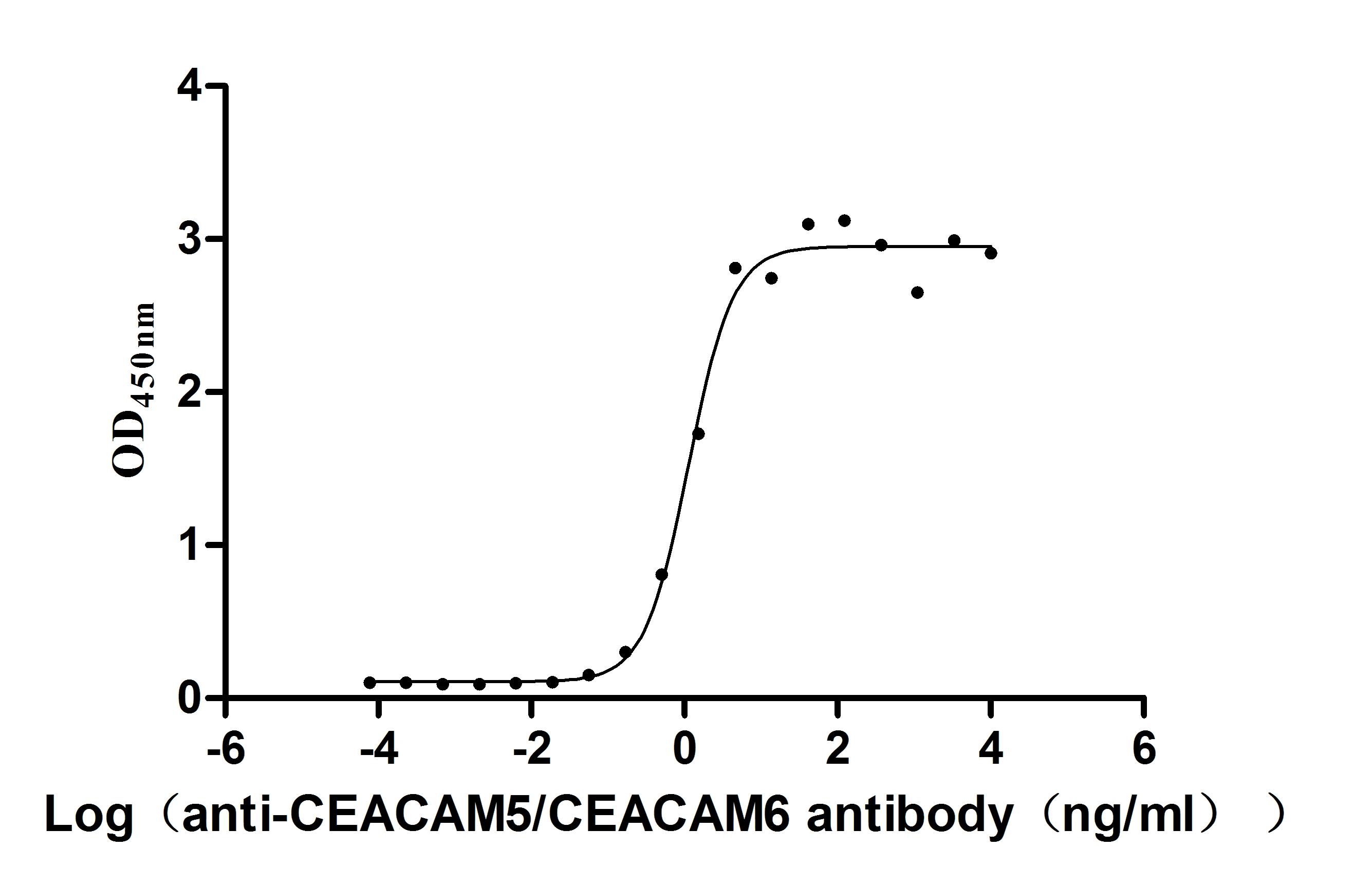
Measured by its binding ability in a functional ELISA. Immobilized Human CEACAM6 (CSB-MP005166HU) at 2 μg/mL can bind Anti- CEACAM5/CEACAM6 recombinant antibody, the EC50 is 0.9430-1.377 ng/mL.
References
[1] Wu, Cheng-Yu, et al. "CEACAM6 as a Novel Therapeutic Target to Boost HO-1-mediated Antioxidant Defense in COPD." American Journal of Respiratory and Critical Care Medicine 207.12 (2023): 1576-1590.
[2] Zang, Mingde, et al. "CEACAM6 promotes tumor angiogenesis and vasculogenic mimicry in gastric cancer via FAK signaling." Biochimica et Biophysica Acta (BBA)-Molecular Basis of Disease 1852.5 (2015): 1020-1028.
[3] Thomas, Jerin, et al. "CEACAMS 1, 5, and 6 in disease and cancer: interactions with pathogens." Genes & Cancer 14 (2023): 12.
[4] Blumenthal, Rosalyn D., et al. "Expression patterns of CEACAM5 and CEACAM6 in primary and metastatic cancers." BMC cancer 7 (2007): 1-15.
[5] Zang, Mingde, et al. "Dual role of carcinoembryonic antigen-related cell adhesion molecule 6 expression in predicting the overall survival of gastric cancer patients." Scientific Reports 7.1 (2017): 10773.
[6] Jantscheff, Peter, et al. "Expression of CEACAM6 in resectable colorectal cancer: a factor of independent prognostic significance." Journal of clinical oncology 21.19 (2003): 3638-3646.
[7] Blumenthal, Rosalyn D., Hans J. Hansen, and David M. Goldenberg. "Inhibition of adhesion, invasion, and metastasis by antibodies targeting CEACAM6 ( NCA-90) and CEACAM5 (Carcinoembryonic Antigen)." Cancer research 65.19 (2005): 8809-8817.
[8] Duxbury, Mark S., et al. "CEACAM6 is a novel biomarker in pancreatic adenocarcinoma and PanIN lesions." Annals of surgery 241.3 (2005): 491.
[9] Blumenthal R D, Leon E, Hansen H J, et al. Expression patterns of CEACAM5 and CEACAM6 in primary and metastatic cancers[J]. BMC cancer, 2007, 7: 1-15.
[10] Johnson, Benny, and Daruka Mahadevan. "Emerging role and targeting of carcinoembryonic antigen-related cell adhesion molecule 6 (CEACAM6) in human malignancies." Clinical cancer drugs 2.2 (2015): 100-111.
[11] Liu, Yingying, et al. "FOXP3 and CEACAM6 expression and T cell infiltration in the occurrence and development of colon cancer." Oncology Letters 11.6 ( 2016): 3693-3701.
[12] Pinkert, Jessica, et al. "T cell-mediated elimination of cancer cells by blocking CEACAM6-CEACAM1 interaction." Oncoimmunology 11.1 ( 2022): 2008110.
[13] Ieta, K., et al. "CEACAM6 gene expression in intrahepatic cholangiocarcinoma." British journal of cancer 95.4 (2006): 532-540.
[14] Blumenthal R D, Hansen H J, Goldenberg D M. Inhibition of adhesion, invasion, and metastasis by antibodies targeting CEACAM6 (NCA-90) and CEACAM5 (Carcinoembryonic Antigen)[J]. Cancer research, 2005, 65(19): 8809-8817.
[15] Chen, Jianmin, et al. "CEACAM6 induces epithelial-mesenchymal transition and mediates invasion and metastasis in pancreatic cancer." International journal of oncology 43.3 (2013): 877-885.
[16] Duxbury, Mark S., et al. "Overexpression of CEACAM6 promotes insulin-like growth factor I-induced pancreatic adenocarcinoma cellular invasiveness. " Oncogene 23.34 (2004): 5834-5842.
[17] Duxbury, Mark S., et al. "A novel role for carcinoembryonic antigen-related cell adhesion molecule 6 as a determinant of gemcitabine chemoresistance in pancreatic adenocarcinoma cells." Cancer research 64.11 (2004): 3987-3993.
[18] Cheng, Tsai-Mu, et al. "Single domain antibody against carcinoembryonic antigen-related cell adhesion molecule 6 (CEACAM6) inhibits proliferation, migration, invasion and angiogenesis of pancreatic cancer cells." European Journal of Cancer 50.4 (2014): 713-721.
[19] Balk-Møller, Emilie, et al. "A marker of endocrine receptor-positive cells, CEACAM6, is shared by two major classes of breast cancer: luminal and HER2- enriched." the american journal of pathology 184.4 (2014): 1198-1208.
[20] Rizeq, Balsam, Zain Zakaria, and Allal Ouhtit. "Towards understanding the mechanisms of actions of carcinoembryonic antigen-related cell adhesion molecule 6 in cancer progression." Cancer science 109.1 (2018): 33-42.
[21] Rodia, Maria Teresa, et al. "LGALS4, CEACAM6, TSPAN8, and COL1A2: Blood markers for colorectal cancer-validation in a cohort of subjects with positive fecal immunochemical test results." Clinical Colorectal Cancer 17.2 (2018): e217-e228.
[22] Jin, Chunhui, et al. "T cell immunity induced by a bivalent Salmonella-based CEACAM6 and 4-1BBL vaccines in a rat colorectal cancer model." Oncology letters 13.5 (2017): 3753-3759.
[23] Duxbury, Mark S., et al. "c-Src-dependent cross-talk between CEACAM6 and αvβ3 integrin enhances pancreatic adenocarcinoma cell adhesion to extracellular matrix components." Biochemical and biophysical research communications 317.1 (2004): 133-141.
[24] Zhang, Yunqiang, et al. "CEACAM6 promotes tumor migration, invasion, and metastasis in gastric cancer." Acta Biochim Biophys Sin 46.4 (2014): 283-290.
[25] Li, Yingmei, et al. "Comprehensive RNA analysis of CSF reveals a role for CEACAM6 in lung cancer leptomeningeal metastases." NPJ Precision Oncology 5.1 ( 2021): 90.
[26] Sheng-lian, L. A. I., M. A. O. Min, and H. U. A. N. G. Lin. "Expression and significance analysis of CEACAM6, miR-146b and S100A6 in thyroid cancer." Journal of Hebei Medical University 44.5 (2023): 531.
[27] Zhu, Rujian, et al. "Carcinoembryonic antigen related cell adhesion molecule 6 promotes the proliferation and migration of renal cancer cells through the ERK/AKT signaling pathway." Translational Andrology and Urology 8.5 (2019): 457.
[28] Kanderová, Veronika, Ondřej Hrušák, and Tomáš Kalina. "Aberrantly expressed CEACAM6 is involved in the signaling leading to apoptosis of acute lymphoblastic leukemia cells." Experimental hematology 38.8 (2010): 653-660.
[29] Lasa, Adriana, et al. "High expression of CEACAM6 and CEACAM8 mRNA in acute lymphoblastic leukemias." Annals of hematology 87 (2008): 205-211.
[30] Steiner, N., et al. "Levels of CEACAM6 in peripheral blood are elevated in patients with plasma cell disorders: a potential new diagnostic marker and a new therapeutic target?." Disease Markers 2019 (2019).
[31] Witzens-Harig, Mathias, et al. "Tumor cells in multiple myeloma patients inhibit myeloma-reactive T cells through carcinoembryonic antigen-related cell adhesion molecule-6." Blood, The Journal of the American Society of Hematology 121.22 (2013): 4493-4503.
[32] Zhang, Feng, et al. "ING5 inhibits cancer aggressiveness via preventing EMT and is a potential prognostic biomarker for lung cancer." Oncotarget 6.18 ( 2015): 16239.
[33] Strickland, Laura A., et al. "Preclinical evaluation of carcinoembryonic cell adhesion molecule (CEACAM) 6 as potential therapy target for pancreatic adenocarcinoma." The Journal of Pathology: A Journal of the Pathological Society of Great Britain and Ireland 218.3 (2009): 380-390.
[34] Riley, Christopher J., et al. "Design and activity of a murine and humanized anti-CEACAM6 single-chain variable fragment in the treatment of pancreatic cancer." Cancer research 69.5 (2009): 1933-1940.
[35] Pandey, Ritu, et al. "Carcinoembryonic antigen cell adhesion molecule 6 (CEACAM6) in Pancreatic Ductal Adenocarcinoma (PDA): an integrative analysis of a novel therapeutic target." Scientific reports 9.1 (2019): 18347.


Comments
Leave a Comment