Call us
301-363-4651 (Available 9 a.m. to 5 p.m. CST from Monday to Friday)
DSG3 is part of the DSGs family (DSG1, DSG2, DSG3, DSG4) within the calcineurin superfamily of cell adhesion molecules. Recent research has highlighted the significance of adhesion molecules in relation to tumors. These molecules are categorized into groups such as the calcineurin superfamily, selectin, immunoglobulin superfamily, integrins, and others. E-cadherin is well-studied for its role in tumor invasion within the calcineurin superfamily. DSG3, a newer adhesion factor, was initially known as an antigen in (Pemphigus Vulgaris, PV), but recent studies have shown its significance in tumors. This suggests that investigating DSG3 further might lead to new diagnostic and therapeutic approaches for diseases related to this protein.
4. The Clinical Potential of DSG3 Targeted Therapy
5. CUSABIO DSG3 Recombinant Proteins & Antibodies for Research Use
Desmoglein 3 (DSG3) is a core protein in the DSGs family, located on human chromosome 18q12.1-q12.2. It plays a key role in forming intercellular junctions and belongs to the calcineurin superfamily of cell adhesion molecules. DSG3 has a molecular mass of about 160 kDa and shares similarities with calcium-dependent cell adhesion molecules. Like typical calcineurin, its structure comprises five extracellular domains (EC1 to EC5), which interact with other cellular proteins to maintain cell-to-cell adhesion and junctions (Figure 1) [1-3].
The current study reveals that DSG3 is exclusively located in the keratinocyte membrane within the epidermal layer of stratified squamous epithelium. Its primary expression occurs in the granular and subgranular layers. DSG3, in conjunction with proteins like DSG1, plays a pivotal role in forming intercellular desmosomes. These desmosomes are crucial for maintaining tissue structure and facilitating strong cell-to-cell connections, ensuring the integrity and stability of the tissue [1-4].
Furthermore, DSG3 is a significant target in Pemphigus Vulgaris (PV), a subtype of pemphigus. In PV patients, the immune system generates anti-DSG3 antibodies that bind to DSG3, disrupting intercellular junctions. This disruption results in the formation of blisters and maculopapular blisters on the skin and mucous membranes. Thus, DSG3 is one of the important factors in the pathogenesis of PV [1-4].
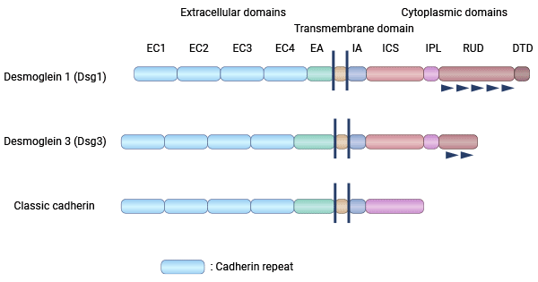
Figure 1. DSG3 structure [3]
The exact mechanisms driving blister formation in pemphigus vulgaris (PV) remain unclear. This process involves a complex interplay of intracellular signaling events (such as Src, p38MAPK, PKC, ERK), factors like Bax, FasL, and Bcl-2, DSG3 degradation, apoptosis, and genetic factors that promote autoantibody production [5-7]. DSG3 is notably involved in maintaining the adhesion between keratinocytes. The binding of specific antibodies to DSG3 disrupts this adhesion—a crucial step in PV's development. However, the precise mechanisms by which DSG3 influences intracellular signaling in PV pathogenesis are not fully understood.
Recent research has identified a serum immunoglobulin in PV patients, PV-IgG, that targets DSG3 and increases p53 protein expression. To confirm this, two experiments were conducted. One used PV patient sera with polyclonal antibodies against DSG3, while the other utilized a monoclonal antibody called AK23, specific to the DSG3 adhesion site. In both cases, exposure to PV serum or AK23 treatment significantly raised p53 expression while decreasing DSG3 protein levels. These results indicate a connection between elevated p53 triggered by PV-IgG or AK23 and specific interference with DSG3. This strongly supports the activation of the DSG3/p53 pathway in PV, suggesting its potential role in the disease's pathogenesis (Figure 2) [5].
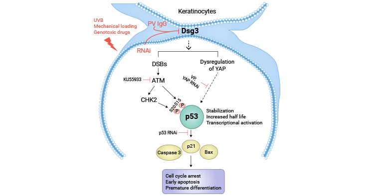
Figure 2. The model for p53 activation in response to DSG3 depletion in keratinocytes [5]
Little is known about the biological functions and mechanistic roles of DSG3 in cancer. A study revealed that in squamous cell carcinoma, DSG3 regulates c-Jun/AP-1 activity and protein kinase C (PKC)-mediated phosphorylation of Ezrin-Thr567, leading to enhanced cancer cell motility and thus promoting cancer cell metastasis [8].
Specifically, DSG3 collaborates with the Ezrin complex at the plasma membrane, crucial for its proper interaction with F-actin and CD44. Knocking down DSG3 disrupts these interactions. Pharmacological inhibitors targeting various serine/threonine kinases (e.g., PKC and Rho kinase) effectively inhibit the increased phosphorylation of Ezrin in DSG3 overexpressing cells. Additionally, DSG3 overexpression leads to a significant increase in c-Jun S63 phosphorylation. Collectively, this study unveils a novel regulatory mechanism involving DSG3, encompassing c-Jun/AP-1 activation and PKC-dependent Ezrin phosphorylation processes, which could contribute to DSG3-associated cancer metastasis (Figure 3) [8-10].
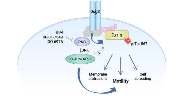
Figure 3. DSG3 promotes tumor via PKC/AP-1 [8]
Pemphigus is a rare skin disease causing recurrent blisters on the skin and mucous membranes. The most common subtype, Pemphigus Vulgaris (PV), starts with low-level autoantibodies and progresses to clinical symptoms. It's classified into four main types: pemphigus vulgaris, pemphigus foliaceus, paraneoplastic pemphigus, and IgA pemphigus. PV is known for its severe clinical symptoms, affecting both the skin and mucous membranes, often leading to complications like hypo-proteinemia, infections, and sepsis. DSG1 and/or DSG3 IgG antibodies are believed to primarily cause PV symptoms [11-13].
Studies show a close connection between DSG3's pathogenicity and PV severity. Anti-DSG3 antibodies are frequently detected in PV patients' serum. PV skin lesion severity is linked to DSG1 antibody levels, while mucosal lesion severity correlates with DSG3 antibody levels. Although current clinical tests use the indirect immunofluorescence (IIF) method to detect circulating antibodies, the specific blister-causing target antigen remains unclear [14-16]. Technological advances highlight DSG1, DSG3, BP180, and BP230 in dermatosis herpetiformis. ELISA, known for its accuracy, is increasingly used to detect DSG3 antibodies. It aids precise pemphigus antibody detection, distinguishes PV from other types, and enhances clinical diagnosis [17-19].
DSG3, the specific antigen of pemphigus vulgaris, has gained attention not only in pemphigus research but also in relation to certain tumors. In studies using the DJM-1 cell line, depletion of "DSG3-depleted bridge particles" led to weakened adhesion between epidermal cells, resulting in skin lesions [20-21]. Furthermore, DSG3 has been implicated in the development of nasopharyngeal carcinoma, with high DSG3 expression considered a key factor promoting the tumor [22]. In the context of human squamous cell carcinoma, reduced intercellular bridge grains are observed, and DSG3 in patient serum serves as a diagnostic indicator [8].
In lung cancer research, among various indicators like CD44 v6, MMPs, TIMPs, p53, VEGF, EGFR, and COX-2, DSG3 has emerged as a valuable prognostic marker. Immunohistochemistry studies link DSG3 expression to tumor type, differentiation degree, and patient survival rates. Positive DSG3 expression is associated with better survival rates, making it a potential prognostic indicator for lung cancer [23-26].
Currently, there is limited research on DSG3, but studies are exploring its potential in pemphigus diagnosis and treatment, including the development of DSG3-targeting drugs for immune and skin disorders. Recent mouse model research demonstrated that Stable/Functional-Induced Regulatory T Cells (S/F-iTregs) can reduce autoimmune markers related to pemphigus [27]. Moreover, S/F-iTregs from aspergillosis patients were found to inhibit T cell proliferation in vitro [27]. Cabaletta Bio, Inc. is in the process of developing a preclinical CAR T immunotherapy known as DSG3+DSG1 CAART for immune aspergillosis. Foreign studies have also highlighted DSG3's role in regulating tumors. Collectively, DSG3, associated with autoimmune pemphigus and tumors, has clinical promise.
DSG3, a member of the DSGs family, belongs to calcineurin superfamily of cell adhesion molecules. It has gained attention for its roles in autoimmune pemphigus, tumor development, and potential therapeutic applications. Studies have explored its significance in pemphigus diagnosis and treatment, including the development of DSG3-targeted drugs for immune and skin disorders. Additionally, DSG3 has been associated with the regulation of tumor growth, indicating its clinical relevance in both autoimmune diseases and cancer.
To fully support researchers and pharmaceutical companies in their research on DSG3 in pemphigus and tumors, CUSABIO presents DSG3 active proteins & antibodies to support your research on the mechanism of DSG3 or its potential clinical value.
CUSABIO DSG3 Protein
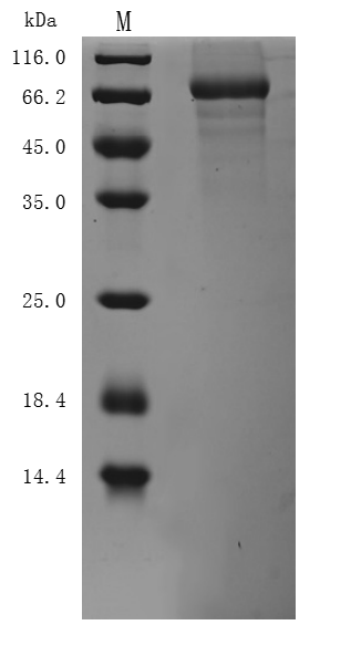
● Recombinant Human Desmoglein-3(DSG3),partial (Active) (Code: CSB-BP007205HUc7)
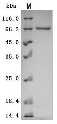
The high purity is greater than 95% as determined by SDS-PAGE.
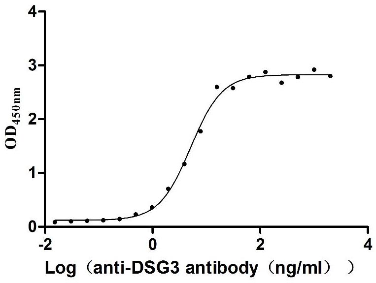
Immobilized Human DSG3 at 2 μg/mL can bind Anti-DSG3 recombinant antibody (CSB-RA007205A0HU), the EC50 is 4.552-5.720 ng/mL.
● Recombinant Mouse Desmoglein-3(Dsg3),partial (Active) (Code: CSB-MP007205MOd7)
The high purity is greater than 85% as determined by SDS-PAGE.
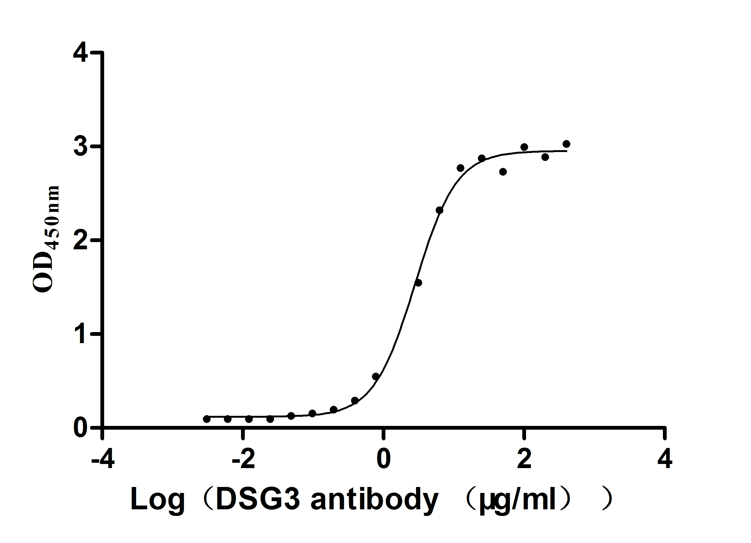
Immobilized Mouse Dsg3 at 5 μg/ml can bind Anti-DSG3 recombinant antibody (CSB-RA007205A0HU), the EC50 is 2.624-3.182 μg/ml.
CUSABIO DSG3 antibody
DSG3 Recombinant Monoclonal Antibody (Code: CSB-RA007205A0HU)
References
[1] Nguyen-Thoi, T., et al. "A cell-based smoothed discrete shear gap method (CS-DSG3) using triangular elements for static and free vibration analyses of shell structures." International Journal of Mechanical Sciences 74 (2013): 32-45.
[2] Ishikawa, H., et al. "Cloning of the mouse desmoglein 3 gene (Dsg3): interspecies conservation within the cadherin superfamily." Experimental dermatology 9.4 (2000): 229-239.
[3] Amagai, Masayuki. "Desmoglein as a target in autoimmunity and infection." Journal of the American Academy of Dermatology 48.2 (2003): 244-252.
[4] Heupel, Wolfgang-Moritz, et al. "Pemphigus vulgaris IgG directly inhibit desmoglein 3-mediated transinteraction." The Journal of Immunology 181.3 (2008): 1825-1834.
[5] Rehman, Ambreen, and Hong Wan. "A novel regulatory pathway of Desmoglein-3 in keratinocyte stress response." Journal of Cellular Signaling 1.4 (2020): 169-179.
[6] Bumiller-Bini Hoch, Valéria, et al. "Marked to Die-Cell Death Mechanisms for Keratinocyte Acantholysis in Pemphigus Diseases." Life 12.3 (2022): 329.
[7] Jolly, Puneet S., et al. "p38MAPK signaling and desmoglein-3 internalization are linked events in pemphigus acantholysis." Journal of Biological Chemistry 285.12 (2010): 8936-8941.
[8] Brown, L., et al. "Desmoglein 3 promotes cancer cell migration and invasion by regulating activator protein 1 and protein kinase C-dependent-Ezrin activation." Oncogene 33.18 (2014): 2363-2374.
[9] Savci-Heijink, Cemile Dilara, et al. "The role of desmoglein-3 in the diagnosis of squamous cell carcinoma of the lung." The American journal of pathology 174.5 (2009): 1629-1637.
[10] Baron, Sylvain, et al. "Unimpaired skin carcinogenesis in Desmoglein 3 knockout mice." PloS one 7.11 (2012): e50024.
[11] Amagai, Masayuki, et al. "Antibodies against desmoglein 3 (pemphigus vulgaris antigen) are present in sera from patients with paraneoplastic pemphigus and cause acantholysis in vivo in neonatal mice." The Journal of clinical investigation 102.4 (1998): 775-782.
[12] Brandão, Maria Luiza Figueiredo Braga, et al. "Refractory pemphigus vulgaris associated with herpes infection: case report and review." Revista do Instituto de Medicina Tropical de São Paulo 53 (2011): 113-117.
[13] Pitoia, Fabián, et al. "Prevalence of thyroid autoimmunity in patients with pemphigus vulgaris." MEDICINA-BUENOS AIRES- 65.4 (2005): 307.
[14] Scully, Crispian, and Stephen J. Challacombe. "Pemphigus vulgaris: update on etiopathogenesis, oral manifestations, and management." Critical Reviews in Oral Biology & Medicine 13.5 (2002): 397-408.
[15] Baker, John, Kristina Seiffert-Sinha, and Animesh A. Sinha. "Case report: Documentation of cutaneous only pemphigus vulgaris without history of mucosal lesions in North America." Frontiers in Immunology 13 (2022): 969279.
[16] Arbache, Samia Trigo, et al. "Immunofluorescence testing in the diagnosis of autoimmune blistering diseases: overview of 10-year experience." Anais brasileiros de dermatologia 89 (2014): 885-889.
[17] Candiz, María Emilia, et al. "Diagnóstico serológico de patologías ampollares autoinmunitarias." Dermatología Argentina 24.4 (2018): 177-184.
[18] Lim, Yen Loo, et al. "Autoimmune Pemphigus: latest advances and emerging therapies." Frontiers in Molecular Biosciences 8 (2022): 808536.
[19] Calle Isaza, Juliana, Isabel Cristina Ávila Gómez, and Ana María Abreu Vélez. "Autoimmune blistering diseases of the pemphigus group." Iatreia 27.3 (2014): 309-319.
[20] Aoyama, Yurni, and Yasuo Kitajima. "Pemphigus vulgaris-IgG causes a rapid depletion of desmoglein 3 (Dsg3) from the Triton X-100 soluble pools, leading to the formation of Dsg3-depleted desmosomes in a human squamous carcinoma cell line, DJM-1 cells." Journal of Investigative Dermatology 112.1 (1999): 67-71.
[21] Tsuchisaka, A., et al. "Presence of autoimmune regulator and absence of desmoglein 1 in a thymoma in a patient with pemphigus foliaceus." British Journal of Dermatology 173.1 (2015): 268-271.
[22] Huang, Yu-Mei, et al. "Integrated analysis of bulk and single-cell RNA sequencing reveals the interaction of PKP1 and tumor-infiltrating B cells and their therapeutic potential for nasopharyngeal carcinoma." Frontiers in Genetics 13 (2022): 935749.
[23] Dong, Boming, et al. "Computed tomographic images reflect the biologic behavior of small lung adenocarcinoma: they correlate with cell proliferation, microvascularization, cell adhesion, degradation of extracellular matrix, and K-ras mutation." The Journal of Thoracic and Cardiovascular Surgery 130.3 (2005): 733-739.
[24] Khoor, A., et al. "DSG3/TTF-1: A single chromogen antibody cocktail to distinguish between Squamous Cell Carcinoma (SCC) and adenocarcinoma of the lung." Virchows Arch 467.1 (2015): S1-S279.
[25] ZHANG, Xindong, et al. "Expression and clinical significance of CK5/6, DSG3, P40, TTF-1, CK7, NapsinA in small biopsy specimens of non-small cell lung cancer." Chinese Journal of Primary Medicine and Pharmacy (2020): 218-221.
[26] Pancewicz, J., and W. Niklinska. "A brief overview of clinical implications of desmoglein 3 in lung cancer." Progress in Health Sciences 11.2 (2021): 141-144.
[27] Collaborators at the Keio and Osaka Universities Present Key Non-Clinical Data with Stable/Functional-Induced Regulatory T Cells (S/F-iTregs) in Autoimmune Disease Models.


Comments
Leave a Comment