ARAF Recombinant Monoclonal Antibody

Code CSB-RA044921A0HU
Size US$210
Order now
Image
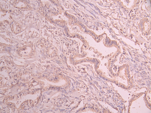
  • IHC image of CSB-RA044921A0HU diluted at 1:100 and staining in paraffin-embedded human cervical cancer performed on a Leica BondTM system. After dewaxing and hydration, antigen retrieval was mediated by high pressure in a citrate buffer (pH 6.0). Section was blocked with 10% normal goat serum 30min at RT. Then primary antibody (1% BSA) was incubated at 4°C overnight. The primary is detected by a Goat anti-rabbit polymer IgG labeled by HRP and visualized using 0.05% DAB.
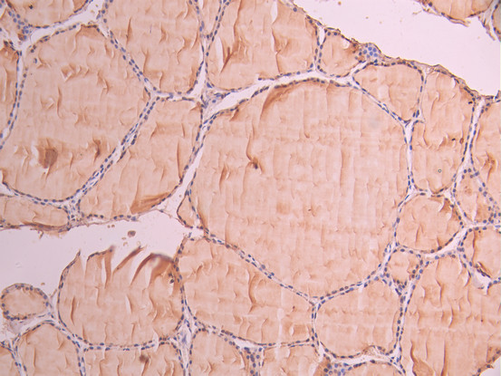
  • IHC image of CSB-RA044921A0HU diluted at 1:100 and staining in paraffin-embedded human thyroid tissue performed on a Leica BondTM system. After dewaxing and hydration, antigen retrieval was mediated by high pressure in a citrate buffer (pH 6.0). Section was blocked with 10% normal goat serum 30min at RT. Then primary antibody (1% BSA) was incubated at 4°C overnight. The primary is detected by a Goat anti-rabbit polymer IgG labeled by HRP and visualized using 0.05% DAB.
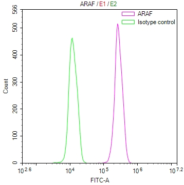
  • Overlay Peak curve showing Hela cells stained with CSB-RA044921A0HU (red line) at 1:100. The cells were fixed in 4% formaldehyde and permeated by 0.2% TritonX-100 for 10min. Then 10% normal goat serum to block non-specific protein-protein interactions followed by the antibody (1ug/1*106cells) for 45min at 4℃. The secondary antibody used was FITC-conjugated goat anti-rabbit IgG (H+L) at 1/200 dilution for 35min at 4℃.Control antibody (green line) was Rabbit IgG (1ug/1*106cells) used under the same conditions. Acquisition of >10,000 events was performed.
Have Questions? Leave a Message or Start an on-line Chat

Product Details

Uniprot No.
Target Names
Alternative Names
Serine/threonine-protein kinase A-Raf (EC 2.7.11.1) (Proto-oncogene A-Raf) (Proto-oncogene A-Raf-1) (Proto-oncogene Pks), ARAF, ARAF1 PKS PKS2
Species Reactivity
Human
Immunogen
A synthesized peptide from human ARAF protein
Immunogen Species
Homo sapiens (Human)
Conjugate
Non-conjugated
Clonality
Monoclonal
Isotype
Rabbit IgG
Clone No.
18D4
Purification Method
Affinity-chromatography
Concentration
It differs from different batches. Please contact us to confirm it.
Buffer
Rabbit IgG in 10mM phosphate buffered saline , pH 7.4, 150mM sodium chloride, 0.05% BSA, 0.02% sodium azide and 50% glycerol.
Form
Liquid
Tested Applications
ELISA, IHC, FC
Recommended Dilution
Application Recommended Dilution
IHC 1:50-1:200
FC 1:50-1:200
Troubleshooting and FAQs
Storage
Upon receipt, store at -20°C or -80°C. Avoid repeated freeze.
Lead Time
Basically, we can dispatch the products out in 1-3 working days after receiving your orders. Delivery time maybe differs from different purchasing way or location, please kindly consult your local distributors for specific delivery time.
Description

ARAF (A-Raf proto-oncogene, serine/threonine kinase) functions as a critical upstream activator in the MAPK/ERK signaling cascade, playing essential roles in cell proliferation, differentiation, and survival. As one of three RAF family kinases, ARAF contributes to signal transduction from cell surface receptors to the nucleus, making it a significant target for researchers investigating oncogenic signaling pathways, developmental biology, and therapeutic resistance mechanisms in cancer.

This recombinant monoclonal antibody, clone 18D4, offers the reproducibility and consistency that demanding research applications require. Generated against a synthetic peptide derived from human ARAF protein, the antibody is produced using recombinant technology, ensuring sequence-defined specificity and eliminating the lot-to-lot variability often encountered with traditional hybridoma-derived antibodies. This consistency is particularly valuable for longitudinal studies or multi-site collaborations where experimental reproducibility is paramount.

Validation testing demonstrates reliable performance across multiple platforms. Immunohistochemistry studies confirm clear detection in paraffin-embedded human cervical cancer and thyroid tissue sections at 1:100 dilution using citrate buffer antigen retrieval, providing researchers flexibility when examining ARAF expression patterns in clinical specimens. Flow cytometry analysis using HeLa cells shows distinct positive signal separation from isotype control, enabling quantitative assessment of ARAF levels in cell populations at dilutions between 1:50 and 1:200.

The unconjugated format and affinity-purified rabbit IgG preparation allow integration into existing detection workflows using standard secondary antibody systems. Supplied in a glycerol-containing buffer for optimal stability, this antibody serves researchers exploring RAF-MEK-ERK pathway dynamics, tumor progression mechanisms, and targeted therapy development.

Usage
For Research Use Only. Not for use in diagnostic or therapeutic procedures.

Customer Reviews and Q&A

 Customer Reviews

There are currently no reviews for this product.

Submit a Review here

Target Background

Function
Involved in the transduction of mitogenic signals from the cell membrane to the nucleus. May also regulate the TOR signaling cascade.; Serves as a positive regulator of myogenic differentiation by inducing cell cycle arrest, the expression of myogenin and other muscle-specific proteins, and myotube formation.
Gene References into Functions
  1. The constitutive or induced re-localization of A-Raf to the plasma membrane compromises its ability to efficiently sequester and inactivate MST2, thus rendering cells susceptible to apoptosis PMID: 26891695
  2. This review discusses the regulation of A-Raf protein expression, and the roles of A-Raf in apoptosis and cancer, with a special focus on its role in resistance to Raf inhibitors. [review] PMID: 26508523
  3. analysis of FGFR2-PPHLN1 fusion and ARAF mutations in intrahepatic cholangiocarcinoma PMID: 25608663
  4. Aberrant expression of A-, B-, and C-RAF, and COT is frequent in PTC; increased expression of COT is correlated with recurrence of PTC. PMID: 25674762
  5. Dimerization of the kinase ARAF promotes MAPK pathway activation and cell migration. PMID: 25097033
  6. The study identifies somatic activating ARAF mutations in Langerhans cell histiocytosis. PMID: 24652991
  7. Galpha12-ARAF-ERK pathway stimulates RFFL transcription through the transcription factor c-Myc. PMID: 24114843
  8. show that Araf antagonizes mesendoderm induction and patterning activity of Nodal/Smad2 signals in vertebrate embryos by directly inhibiting Smad2 signalling PMID: 23591895
  9. study investigated role of ARAF in cancer cell signaling and examined the role of ARAF in mediating paradoxical activation of the MAPK pathway in cells treated with RAF inhibitors; ARAF seems to stabilize BRAF:CRAF complexes in cells treated with RAF inhibitors and regulate cell signaling in a subtle manner to ensure signaling efficiency PMID: 22926515
  10. Ras pathway activation via EGF treatment induced strong binding between B-Raf and C-Raf and a low level of binding between B-Raf and A-Raf. PMID: 23352452
  11. hnRNP H blocks MST2-mediated apoptosis in cancer cells by regulating A-Raf transcription. PMID: 20145135
  12. In a two-hybrid screen of human fetal liver cDNA library, TH1 was detected as a new interaction partner of A-Raf; this specific interaction may have played a critical role in the activation of A-Raf. PMID: 11952167
  13. A-Raf kinase is negatively regulated by trihydrophobin 1 PMID: 14684750
  14. mutation analysis of the conserved regions in the ARAF gene in human colorectal adenocarcinoma PMID: 14688025
  15. A-Raf residues are identified that bind to specific phosphoinositides, possibly as a mechanism to localize the enzyme to particular membrane microdomains rich in these phospholipids. PMID: 15736953
  16. These data reveal that B-RAF is an important mediator of neuronal survival, migration, and dendrite formation and that A-RAF cannot fully compensate for these functions. PMID: 16980614
  17. Positive regulation of A-RAF by phosphorylation of isoform-specific hinge segment and identification of novel phosphorylation sites. PMID: 18662992
  18. A-Raf interacts with MEK1 and activates MEK1 by phosphorylation. PMID: 8621729

Show More

Hide All

Protein Families
Protein kinase superfamily, TKL Ser/Thr protein kinase family, RAF subfamily
Tissue Specificity
Predominantly in urogenital tissues.
Database Links

HGNC: 646

OMIM: 311010

KEGG: hsa:369

STRING: 9606.ENSP00000366244

UniGene: Hs.446641

icon of phone
Call us
301-363-4651 (Available 9 a.m. to 5 p.m. CST from Monday to Friday)

icon of address
Address
No. 269, Shendun 5th Road, Donghu Hi-Tech Development Area, Hubei Province, 430206, P.R.China
icon of social media
Join us with

Subscribe newsletter

Leave a message

* To protect against spam, please pass the CAPTCHA test below.
CAPTCHA verification
© 2007-2026 CUSABIO TECHNOLOGY LLC All rights reserved. 鄂ICP备15011166号-1
Place an order now

I. Product details

*
*
*
*

II. Contact details

*
*

III. Ship To

*
*
*
*
*
*
*

IV. Bill To

*
*
*
*
*
*
*
*