Call us
301-363-4651 (Available 9 a.m. to 5 p.m. CST from Monday to Friday)
SIRT2, a member of the sirtuin family, encodes the NAD+-dependent deacetylase sirtuin 2. Commonly referred to as SIR2-like protein 2, it primarily localizes in the cytoplasm and regulates cellular processes through deacetylation of target proteins such as α-tubulin and p53. Its mechanism involves modulating microtubule dynamics, cell cycle progression, and stress responses, intersecting with pathways like the MAPK and PI3K/Akt signaling cascades.
Aberrant SIRT2 activity is linked to neurodegenerative diseases (e.g., Parkinson’s, Alzheimer’s) and cancer, where it exhibits context-dependent tumor-suppressive or oncogenic roles. Preclinical studies explore small-molecule modulators, including activators and inhibitors, for therapeutic targeting in metabolic disorders and malignancies, though clinical development remains in early stages.
SIRT2 Recombinant Monoclonal Antibody (CSB-RA061500A0HU)
Validated Data
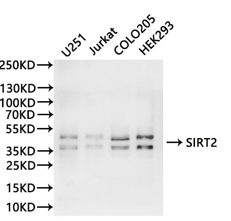
Western Blot
,
Positive WB detected in:U251 whole cell lysate(30µg), Jurkat whole cell lysate(30µg), COLO205 whole cell lysate(30µg), HEK293 whole cell lysate(30µg)
All lanes: SIRT2 antibody at 1:1000
Secondary
Goat polyclonal to rabbit IgG at 1/40000 dilution
Predicted band size: 43 kDa
Observed band size: 36, 43 kDa
Exposure time:120s
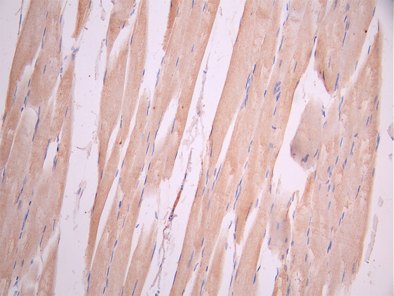
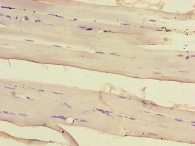
IHC image of CSB-RA061500A0HU diluted at 1:100 and staining in paraffin-embedded human skeletal muscle tissue performed on a Leica BondTM system. After dewaxing and hydration, antigen retrieval was mediated by high pressure in a citrate buffer (pH 6.0). Section was blocked with 10% normal goat serum 30min at RT. Then primary antibody (1% BSA) was incubated at 4°C overnight. The primary is detected by a Goat anti-rabbit polymer IgG labeled by HRP and visualized using 0.05% DAB.
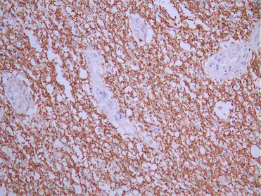
IHC image of CSB-RA061500A0HU diluted at 1:100 and staining in paraffin-embedded human glioma cancer performed on a Leica BondTM system. After dewaxing and hydration, antigen retrieval was mediated by high pressure in a citrate buffer (pH 6.0). Section was blocked with 10% normal goat serum 30min at RT. Then primary antibody (1% BSA) was incubated at 4°C overnight. The primary is detected by a Goat anti-rabbit polymer IgG labeled by HRP and visualized using 0.05% DAB.
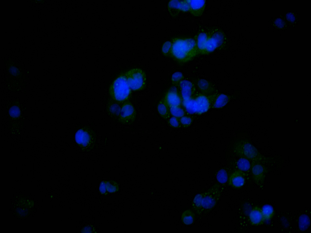
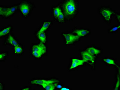
Immunofluorescence staining of HepG2 cell with CSB-RA061500A0HU at 1:50 , counter-stained with DAPI. The cells were fixed in 4% formaldehyde, permeabilized using 0.2% Triton X-100 and blocked in 10% normal Goat Serum. The cells were then incubated with the antibody overnight at 4°C. The secondary antibody was Alexa Fluor 488-congugated AffiniPure Goat Anti-Rabbit IgG(H+L).
SIRT2 Antibody (CSB-PA812889LA01HU)
Validated Data
Immunohistochemistry of paraffin-embedded human skeletal muscle tissue using CSB-PA812889LA01HU at dilution of 1:100
Immunofluorescent analysis of Hela cells using CSB-PA812889LA01HU at dilution of 1:100 and Alexa Fluor 488-congugated AffiniPure Goat Anti-Rabbit IgG(H+L)
The following SIRT2 reagents supplied by CUSABIO are manufactured under a strict quality control system. Multiple applications have been validated and solid technical support is offered.
SIRT2 Antibodies for Homo sapiens (Human)
| Code | Product Name | Species Reactivity | Application |
|---|---|---|---|
| CSB-PA058584 | SIRT2 Antibody | Human | ELISA,WB |
| CSB-PA008284 | SIRT2 Antibody | Human,Mouse,Rat | WB, ELISA |
| CSB-PA157595 | SIRT2 Antibody | Human,Mouse,Rat | ELISA,WB |
| CSB-PA088519 | SIRT2 Antibody | Human,Mouse,Rat | ELISA,WB |
| CSB-PA812889LA01HU | SIRT2 Antibody | Human | ELISA, IHC, IF |
| CSB-PA812889LB01HU | SIRT2 Antibody, HRP conjugated | Human | ELISA |
| CSB-PA812889LC01HU | SIRT2 Antibody, FITC conjugated | Human | |
| CSB-PA812889LD01HU | SIRT2 Antibody, Biotin conjugated | Human | ELISA |
| CSB-RA061500A0HU | SIRT2 Recombinant Monoclonal Antibody | Human | ELISA, WB, IHC, IF |
SIRT2 Proteins for Homo sapiens (Human)
| Code | Product Name | Source |
|---|---|---|
| CSB-YP812889HU CSB-EP812889HU CSB-BP812889HU CSB-MP812889HU CSB-EP812889HU-B |
Recombinant Human NAD-dependent protein deacetylase sirtuin-2 (SIRT2) | Yeast E.coli Baculovirus Mammalian cell In Vivo Biotinylation in E.coli |

