-
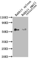
Western Blot
Positive WB detected in: Rabbit spleen tissue, Rabbit small intestine tissue
All lanes: CD14 antibody at 1:2500
Secondary
Goat polyclonal to Mouse IgG at 1/10000 dilution
Predicted band size: 41 kDa
Observed band size: 55 kDa
-
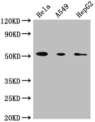
Western Blot
Positive WB detected in: Hela whole cell lysate, A549 whole cell lysate, HepG2 whole cell lysate
All lanes: CD14 antibody at 1:1800
Secondary
Goat polyclonal to Mouse IgG at 1/10000 dilution
Predicted band size: 41 kDa
Observed band size: 55 kDa
-
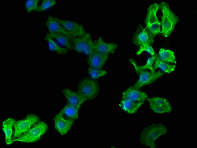
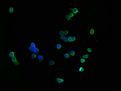
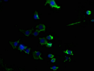
Immunofluorescence staining of A549 cells with CSB-MA004879A0m at 1:90, counter-stained with DAPI. The cells were blocked in 10% normal Goat Serum and then incubated with the primary antibody overnight at 4°C. The secondary antibody was Alexa Fluor 488-congugated AffiniPure Goat Anti-Mouse IgG(H+L).
-
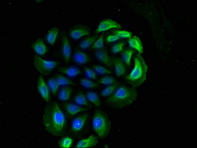
Immunofluorescence staining of Hela cells with CSB-MA004879A0m at 1:90, counter-stained with DAPI. The cells were blocked in 10% normal Goat Serum and then incubated with the primary antibody overnight at 4°C. The secondary antibody was Alexa Fluor 488-congugated AffiniPure Goat Anti-Mouse IgG(H+L).
-
Immunofluorescence staining of HepG2 cells with CSB-MA004879A0m at 1:90, counter-stained with DAPI. The cells were blocked in 10% normal Goat Serum and then incubated with the primary antibody overnight at 4°C. The secondary antibody was Alexa Fluor 488-congugated AffiniPure Goat Anti-Mouse IgG(H+L).
-
Immunofluorescence staining of RAW264.7 cells with CSB-MA004879A0m at 1:90, counter-stained with DAPI. The cells were blocked in 10% normal Goat Serum and then incubated with the primary antibody overnight at 4°C. The secondary antibody was Alexa Fluor 488-congugated AffiniPure Goat Anti-Mouse IgG(H+L).
-
Immunofluorescence staining of SY5Y cells with CSB-MA004879A0m at 1:90, counter-stained with DAPI. The cells were blocked in 10% normal Goat Serum and then incubated with the primary antibody overnight at 4°C. The secondary antibody was Alexa Fluor 488-congugated AffiniPure Goat Anti-Mouse IgG(H+L).
-
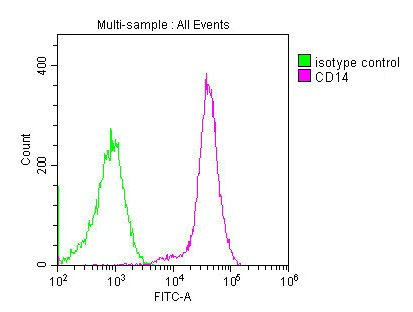
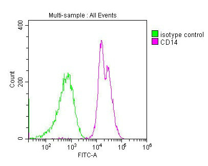
Overlay histogram showing Jurkat cells stained with CSB-MA004879A0m (red line) at 1:180. The cells were incubated in 1x PBS /10% normal goat serum to block non-specific protein-protein interactions followed by primary antibody for 1 h at 4°C. The secondary antibody used was FITC goat anti-mouse IgG(H+L) at 1/200 dilution for 1 h at 4°C. Isotype control antibody (green line) was used under the same conditions. Acquisition of >10,000 events was performed.
-
Overlay histogram showing RAW264.7 cells stained with CSB-MA004879A0m (red line) at 1:180. The cells were incubated in 1x PBS /10% normal goat serum to block non-specific protein-protein interactions followed by primary antibody for 1 h at 4°C. The secondary antibody used was FITC goat anti-mouse IgG(H+L) at 1/200 dilution for 1 h at 4°C. Isotype control antibody (green line) was used under the same conditions. Acquisition of >10,000 events was performed.