
Call us
301-363-4651 (Available 9 a.m. to 5 p.m. CST from Monday to Friday)

A Western blot is only as trustworthy as its normalization. You can capture a crisp band for your target protein, but if the loading control is wrong, the entire quantitative story collapses. Reviewers know this, and so should you.
This guide describes what a loading control antibody is, which experiments require its use, how to choose the right one, and how to troubleshoot common loading control antibody signals.
Table of Contents:
1. What Is A Loading Control Antibody?
2. Which Biological Experiments Require A Loading Control?
3. How to Choose the Right Loading Control Antibody
A loading control antibody is an antibody that specifically detects a loading control protein, a constitutively expressed protein presumed to be present in all samples and whose relative abundance is unaffected by biological variations or experimental conditions [7]. Loading control proteins, typically housekeeping proteins, are routinely used as internal controls for protein loading as well as references in Western blot analysis [2].
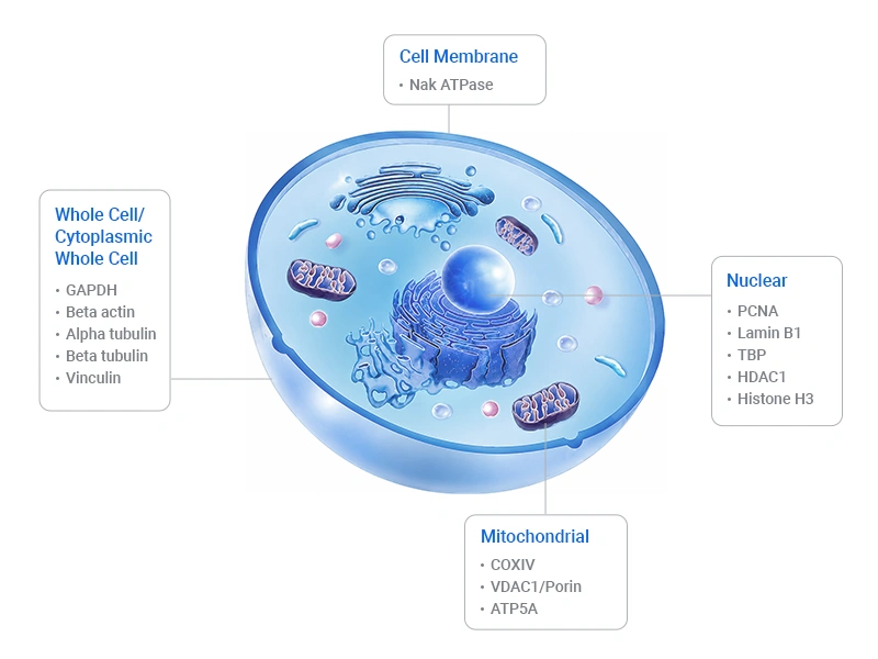
Figure 1. Common loading control proteins
Loading control antibodies serve as internal positive controls. They normalize protein levels between lanes, demonstrate that detection reagents are functioning properly, and confirm that proteins have been efficiently transferred to the membrane [2]. Loading controls can also show whether the edge effect has occurred and allow you to correct for the variation in binding [8].
Quantitative measurements are often subject to error due to overloading the loading control and overreliance on normalization; the burden of proof should lie with the researcher to demonstrate that the chosen loading control reflects quantitative differences in protein concentration [3].
Loading control proteins are constitutively and stably expressed housekeeping proteins used to normalize technical variation across samples, including differences in protein input volume, lysis/extraction efficiency, membrane transfer performance, and detection sensitivity. They are mandatory for all protein-based experiments that perform relative quantification of target protein abundance across multiple experimental groups, as they ensure observed signal differences reflect true biological changes rather than procedural artifacts.
Western blotting is the most canonical application for protein loading controls. Technical variation in protein lysate concentration, pipetting error, uneven electrotransfer, and non-uniform chemiluminescent development can all distort band intensity and be misinterpreted as biological differences in target protein expression.
Journal editors and reviewers routinely require validated loading controls for any quantitative comparison of band intensity between samples. In Western blotting, loading controls compensate for variations in sample loading, electrophoresis, and protein transfer to the membrane. Without a validated loading control, quantitative comparisons of band intensities between samples are scientifically and statistically invalid for peer-reviewed publication [15][12].
In Co-IP and pull-down assays, loading control proteins serve two critical normalization purposes:
EMSA measures DNA-protein binding affinity using nuclear protein extracts. When comparing binding activity across experimental groups, nuclear loading control proteins (such as Histone H3 or Lamin B1) are used to verify equal nuclear protein input and consistent extraction efficiency across samples. This eliminates the possibility that differences in shifted band intensity are due to variable total nuclear protein loading rather than to true changes in transcription factor activity [19]. Input nuclear extracts are routinely validated by Western blot for loading control consistency alongside EMSA experiments.
Dot blot immobilizes protein samples directly on a membrane without electrophoretic separation, making signal intensity highly sensitive to variation in sample volume and concentration. Technical variation from manual spotting introduces even greater bias than gel-based blotting. For quantitative comparisons between sample groups, a validated housekeeping protein loading control is essential to correct for spotting error, following the same normalization principles as traditional western blotting [20][21].
Automated capillary-based Western blot platforms (e.g., Simple Western) perform high-throughput quantitative immunodetection in capillary tubes. The same technical sources of variation (sample input, antibody binding, detection sensitivity) apply, and loading control proteins are equally required for relative quantification of target proteins across experimental groups.
Loading controls are especially critical—and most likely to fail—under the following experimental conditions:
The golden rule: lock down your loading control protein first, then select the corresponding antibody.
This two-step logic prevents the most common mistake: grabbing whichever anti-β-actin is on the shelf.
A valid loading control protein must satisfy four criteria:
The loading control protein must reside in the same subcellular compartment as the site from which your target protein is extracted. Histone H3, for instance, is highly and stably expressed in the nucleus and is not suitable as a reference for cytoplasmic proteins. β-actin, GAPDH, and α-Tubulin are expressed in the cytoplasm and should be selected as loading controls for cytoplasmic proteins. Normalizing a nuclear target against a cytoplasmic control introduces systematic error and invalidates fractionation experiments [3].
Expression Site of common loading control proteins:
| Sample Type | Recommended Loading Control Protein | MW (kDa) | Corresponding Loading Control Antibody |
|---|---|---|---|
| Whole cell / Cytoplasmic | Vinculin (VCL) | 116 | Vinculin (VCL) antibody |
| α-Tubulin | 55 | TUBA1A Monoclonal Antibody | |
| β-Tubulin (TUBB) | 50–55 | TUBB Monoclonal Antibody | |
| β-Actin (ACTB) | 42 | ACTB Monoclonal Antibody | |
| GAPDH | 36 | GAPDH Monoclonal Antibody | |
| Nuclear | Lamin B1 (LMNB1) | 66 | LMNB1 Monoclonal Antibody |
| HDAC1 | 55 | HDAC1 Recombinant Monoclonal Antibody | |
| TBP | 38 | TBP Recombinant Monoclonal Antibody | |
| PCNA | 30 | PCNA Monoclonal Antibody | |
| Histone H3 | 15 | HistoneH3 Monoclonal Antibody | |
| Mitochondrial | VDAC1 | 35 | VDAC1 Recombinant Monoclonal Antibody |
| COX IV | 18 | COX4I1 Monoclonal Antibody | |
| ATP5A | 60 | ATP5A1 Recombinant Monoclonal Antibody | |
| Plasma membrane | Na⁺/K⁺-ATPase | 112 | ATP1A1 Recombinant Monoclonal Antibody |
| Serum | Transferrin | 77 | Transferrin (TF) Recombinant Monoclonal Antibody |
* Molecular weights are approximate and species-dependent. This table reflects consensus practice; each entry must be independently validated for the experimental system in question.
Choose a loading control protein with a molecular weight that differs from your protein of interest, so the bands can be easily distinguished. If these proteins are nearly the same size, band visualization and data interpretation will be compromised.
For clear band resolution, choose a loading control protein with a molecular weight of at least 5 kDa different from your target protein—ideally 20 kDa or more. For example, if your target runs at 42 kDa, β-Actin (42 kDa) will overlap completely; switch to GAPDH (37 kDa) or β-Tubulin (50 kDa) for clean separation [3].
This is the most critical and most often skipped step. Putative housekeeping genes exhibit significant variation in both mRNA and protein content; blind selection of internal controls can result in spurious results [11]. No single gene among the commonly used housekeeping genes can serve as a universal reference across all tissue types under all conditions [12].
A modern approach is to use the GEO database and the web tool GEO2R to rapidly exclude unstable housekeeping genes before committing to an antibody-based loading control. In ischemic heart tissues, actin and tubulin changed significantly, whereas no statistically significant changes were observed in the expression of genes relative to GAPDH. However, unstable housekeeping genes were also found in other animal models of cardiovascular medicine [1].
Common mismatches to avoid:
Housekeeping proteins are highly abundant, and their bands easily saturate at sample loads optimized for low-abundance targets, creating a false "all lanes look equal" signal [22, 2]. To ensure quantitative accuracy:
Once you've locked down the loading control protein, you need an antibody that specifically and reliably detects it.
To achieve reproducible Western blot results, the primary antibody species, isotype, source, catalog and lot numbers, dilution, and incubation conditions must all be provided [7]. Trial experiments aiming to generate a standard curve are recommended when new antibodies or methods are employed [8].
Before committing to a loading control antibody, run a pilot:
Despite their name, housekeeping proteins are not universally constant. Understanding when and why they fail is essential for designing defensible experiments.
The canonical housekeeping proteins are not universally constant. In a developmental time-course of rat retina, β-actin, cyclophilin B, α-tubulin, and lamin A/C all showed significant inter-stage variation, whereas MAPK1 remained stable. Similarly, GAPDH and β-actin protein levels decrease with aging in human skeletal muscle, rendering them unsuitable as loading controls in sarcopenia or myopathy studies [13]. In ischemic cardiac tissue, actin and tubulin varied significantly while GAPDH did not—but in other cardiovascular models, GAPDH itself was among the unstable housekeeping genes.
These findings collectively invalidate the heuristic that "any housekeeper will do." The appropriate control is condition-specific, and its stability must be demonstrated rather than assumed.
Recognition that loading control proteins are rarely in the same linear detection range as the protein of interest means that immunodetection of these proteins often falls outside that range, rendering accurate quantitation impossible [2]. Housekeeping proteins are high-abundance proteins and therefore are often overloaded, particularly when large amounts of total protein are loaded to detect low-abundance target proteins [6].
Over the past decade, using total protein staining instead of a single-protein loading control has become a more acceptable practice. Total protein quantification by Ponceau S, Coomassie Brilliant Blue, and Stain-Free methods has been shown to have advantages over housekeeping proteins for normalization of Western blots—mainly because this normalization does not depend on the expression of a single protein [1].
Common loading controls include housekeeping proteins, such as β-actin or GAPDH, quantified by Western blot, or total protein, quantified using a stain such as Coomassie Brilliant Blue or Ponceau S [10]. A more recently developed method for total protein quantification utilizes stain-free technology, which has a linear dynamic detection range and allows for protein detection on both gels and membranes [14].
Methods include:
Total protein staining does not replace loading control antibodies in every scenario, but it should be part of your validation toolkit—especially when working with precious samples, body fluids, or extreme treatment conditions. The combination of a housekeeping protein with total protein normalization provides redundancy as a strength for higher confidence [6].
Even with the right antibody, Western blot execution can introduce artifacts. Here's how to diagnose and fix the most frequent problems.
Possible causes:
Fixes:
Why it happens: Housekeeping proteins are abundant. At typical loading amounts, their bands are often already saturated, placing them outside the linear detection range and making quantification impossible. Oversaturated exposure of blots should be avoided to maintain the band signal intensity in the linear range for quantitation [7]. Oversaturated bands may mask or at least reduce the differences among samples [6].
Fixes:
Common reasons:
Fixes:
Loading control antibodies remain the most widely used and practical tool for quantitative Western blot normalization and experimental quality control. Choosing a loading control antibody demands evidence, not habit. Their reliability does not depend on a universal “gold standard” protein, but on careful selection matched to subcellular compartment, molecular weight, experimental context, and detection linearity. By treating loading control selection as a deliberate, evidence-based step rather than a default checkbox, you elevate the rigor of your Western blot data and build a foundation that survives peer review.
References:
[1] Wang Q, Han W, Ma C, Wang T, Zhong J. Western blot normalization: Time to choose a proper loading control seriously [J]. Electrophoresis. 2023 May;44(9-10):854-863.
[2] McDonough, A. A., Veiras, L. C., Minas, J. N., & Ralph, D. L. (2014). Considerations when quantitating protein abundance by immunoblot [J]. American Journal of Physiology - Cell Physiology, 308(6), C426.
[3] Aldridge, G. M., Podrebarac, D. M., Greenough, W. T., & Weiler, I. J. (2008). The use of total protein stains as loading controls: An alternative to high-abundance single protein controls in semi-quantitative immunoblotting [J]. Journal of Neuroscience Methods, 172(2), 250.
[4] Gilda JE, Gomes AV. Stain-Free total protein staining is a superior loading control to β-actin for Western blots [J]. Anal Biochem. 2013 Sep 15;440(2):186-8.
[5] Hagner-McWhirter Å, Laurin Y, Larsson A, Bjerneld EJ, Rönn O. Cy5 total protein normalization in Western blot analysis [J]. Anal Biochem. 2015 Oct 1;486:54-61.
[6] Ghosh, R., Gilda, J. E., & Gomes, A. V. (2014). The necessity of and strategies for improving confidence in the accuracy of western blots [J]. Expert Review of Proteomics, 11(5), 549.
[7] Tie, L., Xiao, H., Wu, D. L., Yang, Y., & Wang, P. (2020). A brief guide to good practices in pharmacological experiments: Western blotting [J]. Acta Pharmacologica Sinica, 42(7), 1015.
[8] Taylor, S. C., & Posch, A. (2014). The Design of a Quantitative Western Blot Experiment [J]. BioMed Research International, 2014, 361590.
[9] Romero-Calvo I, Ocón B, et al. Reversible Ponceau staining as a loading control alternative to actin in Western blots [J]. Anal Biochem. 2010 Jun 15;401(2):318-20.
[10] Welinder C, Ekblad L. Coomassie staining as loading control in Western blot analysis [J]. J Proteome Res. 2011 Mar 4;10(3):1416-9.
[11] Rocha-Martins, M., Njaine, B., & Silveira, M. S. (2012). Avoiding Pitfalls of Internal Controls: Validation of Reference Genes for Analysis by qRT-PCR and Western Blot throughout Rat Retinal Development [J]. PLoS ONE, 7(8), e43028.
[12] Li, R., & Shen, Y. (2013). An old method facing a new challenge: Re-visiting housekeeping proteins as internal reference control for neuroscience research [J]. Life Sciences, 92(13), 747.
[13] Vigelsø A, Dybboe R, Hansen CN, Dela F, Helge JW, Guadalupe Grau A. GAPDH and β-actin protein decreases with aging, making Stain-Free technology a superior loading control in Western blotting of human skeletal muscle [J]. J Appl Physiol (1985). 2015 Feb 1;118(3):386-94.
[14] Gilda JE, Gomes AV. Western blotting using in-gel protein labeling as a normalization control: stain-free technology [J]. Methods Mol Biol. 2015;1295:381-91.
[15] Neris RLS, Dobles AMC, Gomes AV. Western Blotting Using In-Gel Protein Labeling as a Normalization Control: Advantages of Stain-Free Technology [J]. Methods Mol Biol. 2021;2261:443-456.
[16] Orantos-Aguilera Y, Sanchez-Lopez I, et al. STIM1-containing contact sites promote direct calcium flux from the endoplasmic reticulum to mitochondria [J]. EMBO J. 2026 Mar;45(5):1760-1793.
[17] Rahman K, Coomer CA, Majdoul S, Ding SY, Padilla-Parra S, Compton AA. Homology-guided identification of a conserved motif linking the antiviral functions of IFITM3 to its oligomeric state [J]. Elife. 2020 Oct 28;9:e58537.
[18] Rogstad, S. M., Sorkina, T., Sorkin, A., & Wu, C. C. (2013). Improved Precision of Proteomic Measurements in Immunoprecipitation Based Purifications Using Relative Quantitation [J]. Analytical Chemistry, 85(9), 4301.
[19] Jelinic P, Eccles LA, et al. The EMSY threonine 207 phospho-site is required for EMSYdriven suppression of DNA damage repair [J]. Oncotarget. 2017 Feb 21;8(8):13792-13804.
[20] Colella AD, Chegenii N, Tea MN, Gibbins IL, Williams KA, Chataway TK. Comparison of Stain-Free gels with traditional immunoblot loading control methodology [J]. Anal Biochem. 2012 Nov 15;430(2):108-10.
[21] Wang JL, Li MQ, Zhang JJ, Xu CJ. Total protein staining with Congo red as an alternative loading control for western blot analysis [J]. Biotech Histochem. 2022 Aug;97(6):404-414.
[22] Dittmer A, Dittmer J. Beta-actin is not a reliable loading control in Western blot analysis. Electrophoresis [J]. 2006 Jul;27(14):2844-5.
[23] Taylor SC, Berkelman T, Yadav G, Hammond M. A defined methodology for reliable quantification of Western blot data [J]. Mol Biotechnol. 2013 Nov;55(3):217-26.




Comments
Leave a Comment