Call us
301-363-4651 (Available 9 a.m. to 5 p.m. CST from Monday to Friday)
AKT1, or protein kinase Bα, is a serine/threonine kinase that plays a key role in intracellular signalling.AKT1 is widely present in a wide range of tissues and cell types and is a central component of the PI3K/AKT signalling pathway. Its mechanism of action is through the recruitment of phosphatidylinositol-3,4,5-trisphosphate (PIP3) produced by PI3K (phosphatidylinositol 3 kinase), which is then activated. Activated AKT1 phosphorylates a variety of substrate proteins and regulates cell growth, survival, metabolism, and apoptosis.AKT1 is involved in signaling pathways including the PI3K/AKT/mTOR pathway, which plays an essential role in cell proliferation, protein synthesis, cell metabolism, and cell survival.AKT1 has also been implicated in cell-cycle regulation, cellular stress response, and cell migration, among other processes. processes. In a biological sense, AKT1 is essential for maintaining the stability of the intracellular environment and adapting to external changes. Abnormalities in its function have been associated with a variety of diseases, especially with the development of tumours, and it is a potential target in cancer therapy.
Recombinant Rat RAC-alpha serine/threonine-protein kinase (Akt1) (CSB-EP001553RA)
Validated Data
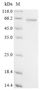
(Tris-Glycine gel) Discontinuous SDS-PAGE (reduced) with 5% enrichment gel and 15% separation gel.
AKT1 Recombinant Monoclonal Antibody (CSB-RA917625A0HU)
Validated Data
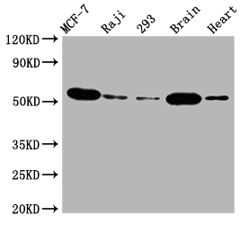
Western Blot
Positive WB detected in: MCF-7 whole cell lysate, Raji whole cell lysate, 293 whole cell lysate, Mouse brain tissue, Rat heart tissue
All lanes: AKT1 antibody at 1:2000
Secondary
Goat polyclonal to rabbit IgG at 1/50000 dilution
Predicted band size: 56 kDa
Observed band size: 56 kDa
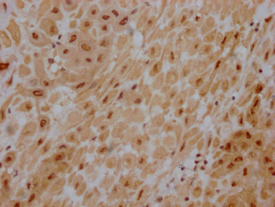
IHC image of CSB-RA917625A0HU diluted at 1:100 and staining in paraffin-embedded human placenta tissue performed on a Leica BondTM system. After dewaxing and hydration, antigen retrieval was mediated by high pressure in a citrate buffer (pH 6.0). Section was blocked with 10% normal goat serum 30min at RT. Then primary antibody (1% BSA) was incubated at 4℃ overnight. The primary is detected by a Goat anti-rabbit IgG polymer labeled by HRP and visualized using 0.05% DAB.
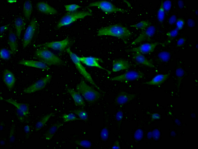
Immunofluorescence staining of Hela Cells with CSB-RA917625A0HU at 1:50, counter-stained with DAPI. The cells were fixed in 4% formaldehyde, permeated by 0.2% TritonX-100, and blocked in 10% normal Goat Serum. The cells were then incubated with the antibody overnight at 4℃. Nuclear DNA was labeled in blue with DAPI. The secondary antibody was FITC-conjugated AffiniPure Goat Anti-Rabbit IgG (H+L).
The following AKT1 reagents supplied by CUSABIO are manufactured under a strict quality control system. Multiple applications have been validated and solid technical support is offered.
AKT1 Antibodies for Homo sapiens (Human)
| Code | Product Name | Species Reactivity | Application |
|---|---|---|---|
| CSB-PA001553GA01HU | AKT1 Antibody | Human | ELISA,WB |
| CSB-PA15905D0Rb | AKT1 Antibody, Biotin conjugated | Human | ELISA |
| CSB-PA15905B0Rb | AKT1 Antibody, HRP conjugated | Human | ELISA |
| CSB-PA15905C0Rb | AKT1 Antibody, FITC conjugated | Human | |
| CSB-PA573922 | Phospho-AKT1 (Ser473) Antibody | Human,Mouse,Rat | ELISA,WB,IHC |
| CSB-PA010227 | Phospho-AKT1 (Thr308) Antibody | Human,Mouse,Rat | ELISA,WB,IHC,IF |
| CSB-PA237749 | Phospho-AKT1 (Thr450) Antibody | Human,Mouse,Rat | ELISA,WB,IHC,IF |
| CSB-PA063905 | AKT1 (Ab-473) Antibody | Human,Mouse,Rat | ELISA,WB,IHC |
| CSB-PA288499 | AKT1 (Ab-308) Antibody | Human,Mouse,Rat | ELISA,WB,IHC |
| CSB-PA969941 | AKT1 (Ab-450) Antibody | Human,Mouse,Rat | ELISA,WB,IHC,IF |
| CSB-PA919481 | AKT1 (Ab-124) Antibody | Human,Mouse,Rat | ELISA,WB |
| CSB-PA996941 | AKT1 (Ab-129) Antibody | Human,Mouse,Rat | ELISA,WB,IHC,IF |
| CSB-PA927050 | AKT1 (Ab-326) Antibody | Human,Mouse,Rat | ELISA,WB,IHC,IF |
| CSB-PA932880 | AKT1/AKT3 (Ab-437/434) Antibody | Human,Mouse | ELISA,WB |
| CSB-PA115404 | Phospho-AKT1 (Ser129) Antibody | Human,Mouse,Rat | ELISA,WB |
| CSB-PA149203 | AKT1 Antibody | Human,Mouse,Rat | ELISA,WB |
| CSB-PA204523 | AKT1 Antibody | Human,Mouse,Rat | ELISA,IHC |
| CSB-PA038073 | Phospho-AKT1/AKT3 (Tyr437/434) Antibody | Human,Mouse,Rat | ELISA,WB,IHC |
| CSB-PA063650 | Phospho-AKT1 (Tyr326) Antibody | Human,Mouse,Rat | ELISA,WB |
| CSB-PA000465 | Phospho-AKT1 (S246) Antibody | Human,Mouse,Rat | WB, IHC, IF, ELISA |
| CSB-PA000468 | Phospho-AKT1 (T450) Antibody | Human,Mouse,Rat | WB, IHC, ELISA |
| CSB-PA000645 | Phospho-AKT1 (S129) Antibody | Human,Mouse,Rat | WB, ELISA |
| CSB-PA000649 | Phospho-AKT1 (Y326) Antibody | Human,Mouse,Rat | WB, ELISA |
| CSB-PA000657 | Phospho-AKT1 (T308) Antibody | Human,Mouse,Rat | WB, ELISA |
| CSB-PA000704 | Phospho-AKT1 (Y474) Antibody | Human,Mouse,Rat,Monkey | WB, IHC, ELISA |
| CSB-PA000732 | Phospho-AKT1 (S473) Antibody | Human,Mouse,Rat | WB, IHC, IF, ELISA |
| CSB-PA000849 | AKT1 Antibody | Human,Mouse,Rat | WB, IHC, ELISA |
| CSB-PA000851 | AKT1 Antibody | Human,Mouse,Rat,Monkey | WB, IHC, IF, ELISA |
| CSB-PA000852 | AKT1 Antibody | Human,Mouse,Rat | WB, IHC, IF, ELISA |
| CSB-PA000855 | AKT1 Antibody | Human,Mouse,Rat | WB, IHC, ELISA |
| CSB-PA008118 | Phospho-AKT1 (S124) Antibody | Human,Mouse,Rat | WB, IHC, ELISA |
| CSB-PA008120 | Phospho-AKT1 (T72) Antibody | Human,Mouse,Rat | WB, IHC, ELISA |
| CSB-PA008122 | AKT1 Antibody | Human,Mouse,Rat | IHC, ELISA |
| CSB-PA080256 | AKT1 Antibody | Human,Mouse,Rat | WB |
| CSB-PA098004 | AKT1 Antibody | Human,Mouse,Rat | ELISA,IHC |
| CSB-PA721992 | AKT1 Antibody | Human,Mouse,Rat | ELISA,WB |
| CSB-PA942120 | AKT1 Antibody | Human,Mouse,Rat | ELISA,WB |
| CSB-PA15905A0Rb | AKT1 Antibody | Human | ELISA, WB, IHC, IF, IP |
| CSB-RA001553A450phHU | Phospho-AKT1 (T450) Recombinant Monoclonal Antibody | Human | ELISA, WB |
| CSB-RA001553A473phHU | Phospho-AKT1 (Ser473) Recombinant Monoclonal Antibody | Human | ELISA, WB, IHC, IF, IP |
| CSB-RA917625A0HU | AKT1 Recombinant Monoclonal Antibody | Human, Mouse, Rat | ELISA, WB, IHC, IF |
AKT1 Antibodies for Arabidopsis thaliana (Mouse-ear cress)
| Code | Product Name | Species Reactivity | Application |
|---|---|---|---|
| CSB-PA655043XA01DOA | AKT1 Antibody | Arabidopsis thaliana | ELISA, WB (ensure identification of antigen) |
AKT1 Antibodies for Drosophila melanogaster (Fruit fly)
| Code | Product Name | Species Reactivity | Application |
|---|---|---|---|
| CSB-PA818217XA01DLU | Akt1 Antibody | Drosophila melanogaster (Fruit fly) | ELISA, WB (ensure identification of antigen) |
AKT1 Antibodies for Oryza sativa subsp. indica (Rice)
| Code | Product Name | Species Reactivity | Application |
|---|---|---|---|
| CSB-PA315034XA01OFF | AKT1 Antibody | Oryza sativa subsp. indica (Rice) | ELISA, WB (ensure identification of antigen) |
AKT1 Antibodies for Oryza sativa subsp. japonica (Rice)
| Code | Product Name | Species Reactivity | Application |
|---|---|---|---|
| CSB-PA612580XA01OFG | AKT1 Antibody | Oryza sativa subsp. japonica (Rice) | ELISA, WB (ensure identification of antigen) |
AKT1 Proteins for Homo sapiens (Human)
| Code | Product Name | Source |
|---|---|---|
| CSB-YP001553HU CSB-BP001553HU CSB-MP001553HU CSB-EP001553HU-B |
Recombinant Human RAC-alpha serine/threonine-protein kinase (AKT1) | Yeast Baculovirus Mammalian cell In Vivo Biotinylation in E.coli |
| CSB-EP001553HU | Recombinant Human RAC-alpha serine/threonine-protein kinase (AKT1) | E.coli |
AKT1 Proteins for Mus musculus (Mouse)
| Code | Product Name | Source |
|---|---|---|
| CSB-YP001553MO CSB-EP001553MO CSB-BP001553MO CSB-MP001553MO CSB-EP001553MO-B |
Recombinant Mouse RAC-alpha serine/threonine-protein kinase (Akt1) | Yeast E.coli Baculovirus Mammalian cell In Vivo Biotinylation in E.coli |
AKT1 Proteins for Rattus norvegicus (Rat)
| Code | Product Name | Source |
|---|---|---|
| CSB-YP001553RA CSB-BP001553RA CSB-MP001553RA CSB-EP001553RA-B |
Recombinant Rat RAC-alpha serine/threonine-protein kinase (Akt1) | Yeast Baculovirus Mammalian cell In Vivo Biotinylation in E.coli |
AKT1 Proteins for Drosophila melanogaster (Fruit fly)
| Code | Product Name | Source |
|---|---|---|
| CSB-YP818217DLU CSB-EP818217DLU CSB-BP818217DLU CSB-MP818217DLU CSB-EP818217DLU-B |
Recombinant Drosophila melanogaster RAC serine/threonine-protein kinase (Akt1), partial | Yeast E.coli Baculovirus Mammalian cell In Vivo Biotinylation in E.coli |
AKT1 Proteins for Xenopus laevis (African clawed frog)
| Code | Product Name | Source |
|---|---|---|
| CSB-YP857839XBE CSB-EP857839XBE CSB-BP857839XBE CSB-MP857839XBE CSB-EP857839XBE-B |
Recombinant Xenopus laevis RAC-alpha serine/threonine-protein kinase (akt1) | Yeast E.coli Baculovirus Mammalian cell In Vivo Biotinylation in E.coli |
AKT1 Proteins for Oryza sativa subsp. japonica (Rice)
| Code | Product Name | Source |
|---|---|---|
| CSB-YP612580OFG CSB-EP612580OFG CSB-BP612580OFG CSB-MP612580OFG CSB-EP612580OFG-B |
Recombinant Oryza sativa subsp. japonica Potassium channel AKT1 (AKT1), partial | Yeast E.coli Baculovirus Mammalian cell In Vivo Biotinylation in E.coli |
AKT1 Proteins for Arabidopsis thaliana (Mouse-ear cress)
| Code | Product Name | Source |
|---|---|---|
| CSB-YP655043DOA CSB-EP655043DOA CSB-BP655043DOA CSB-MP655043DOA CSB-EP655043DOA-B |
Recombinant Arabidopsis thaliana Potassium channel AKT1 (AKT1), partial | Yeast E.coli Baculovirus Mammalian cell In Vivo Biotinylation in E.coli |
AKT1 Proteins for Oryza sativa subsp. indica (Rice)
| Code | Product Name | Source |
|---|---|---|
| CSB-YP315034OFF CSB-EP315034OFF CSB-BP315034OFF CSB-MP315034OFF CSB-EP315034OFF-B |
Recombinant Oryza sativa subsp. indica Potassium channel AKT1 (AKT1), partial | Yeast E.coli Baculovirus Mammalian cell In Vivo Biotinylation in E.coli |
AKT1 ELISA Kit for Homo sapiens (Human)
| Code | Product Name | Sample Types | Sensitivity |
|---|---|---|---|
| CSB-EL001553HU | Human RAC-alpha serine/threonine-protein kinase(AKT1) ELISA kit | serum, plasma, cell lysates | 0.078 ng/mL |

