Call us
301-363-4651 (Available 9 a.m. to 5 p.m. CST from Monday to Friday)
CAP1, also known as Adenylyl cyclase-associated protein 1, is a well - studied protein. It is commonly referred to as CAP. This protein is involved in regulating the actin cytoskeleton. It binds to G - actin and promotes nucleotide exchange, influencing the dynamics of actin polymerization and depolymerization.
CAP1 is associated with the Wnt signaling pathway, which is crucial for cell proliferation and differentiation. Aberrant expression of CAP1 has been linked to various diseases, such as certain types of cancer. Currently, research is underway to develop drugs that target CAP1 to interfere with its function and potentially treat related diseases.
Recombinant Human Adenylyl cyclase-associated protein 1 (CAP1) (CSB-EP004486HUc7)
Validated Data
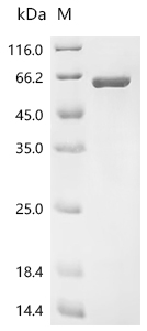
(Tris-Glycine gel) Discontinuous SDS-PAGE (reduced) with 5% enrichment gel and 15% separation gel.
CAP1 Antibody (CSB-PA004486LA01HU)
Validated Data
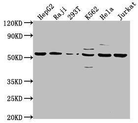
Western Blot
Positive WB detected in: HepG2 whole cell lysate, Raji whole cell lysate, 293T whole cell lysate, K562 whole cell lysate, Hela whole cell lysate, Jurkat whole cell lysate
All lanes: CAP1 antibody at 1:2000
Secondary
Goat polyclonal to rabbit IgG at 1/50000 dilution
Predicted band size: 52 kDa
Observed band size: 52 kDa
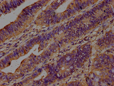
IHC image of CSB-PA004486LA01HU diluted at 1:200 and staining in paraffin-embedded human colon cancer performed on a Leica BondTM system. After dewaxing and hydration, antigen retrieval was mediated by high pressure in a citrate buffer (pH 6.0). Section was blocked with 10% normal goat serum 30min at RT. Then primary antibody (1% BSA) was incubated at 4°C overnight. The primary is detected by a biotinylated secondary antibody and visualized using an HRP conjugated SP system.
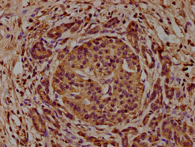
IHC image of CSB-PA004486LA01HU diluted at 1:200 and staining in paraffin-embedded human pancreatic cancer performed on a Leica BondTM system. After dewaxing and hydration, antigen retrieval was mediated by high pressure in a citrate buffer (pH 6.0). Section was blocked with 10% normal goat serum 30min at RT. Then primary antibody (1% BSA) was incubated at 4°C overnight. The primary is detected by a biotinylated secondary antibody and visualized using an HRP conjugated SP system.
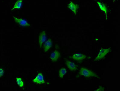
Immunofluorescence staining of U251 cells with CSB-PA004486LA01HU at 1:100, counter-stained with DAPI. The cells were fixed in 4% formaldehyde, permeabilized using 0.2% Triton X-100 and blocked in 10% normal Goat Serum. The cells were then incubated with the antibody overnight at 4°C. The secondary antibody was Alexa Fluor 488-congugated AffiniPure Goat Anti-Rabbit IgG(H+L).
Human Adenylyl cyclase-associated protein 1(CAP1) ELISA kit (CSB-EL004486HU)
Validated Data
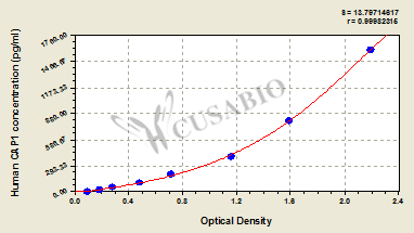
Code: CSB-EL004486HU
Size: 96T,5×96T,10×96T
Sensitivity: 6.25 pg/mL
Detection Range: 25 pg/mL-1600 pg/mL
These standard curves are provided for demonstration only. A standard curve should be generated for each set of samples assayed.
The following CAP1 reagents supplied by CUSABIO are manufactured under a strict quality control system. Multiple applications have been validated and solid technical support is offered.
CAP1 Antibodies for Homo sapiens (Human)
| Code | Product Name | Species Reactivity | Application |
|---|---|---|---|
| CSB-PA004486GA01HU | CAP1 Antibody | Human,Mouse,Rat | ELISA,WB |
| CSB-PA256310 | CAP1 Antibody | Human,Mouse,Rat | ELISA,WB,IHC |
| CSB-PA147089 | CAP1 Antibody | Human,Mouse,Rat | ELISA,WB,IHC |
| CSB-PA004486LA01HU | CAP1 Antibody | Human | ELISA, WB, IHC, IF |
CAP1 Proteins for Homo sapiens (Human)
| Code | Product Name | Source |
|---|---|---|
| CSB-YP004486HU CSB-EP004486HU CSB-BP004486HU CSB-MP004486HU CSB-EP004486HU-B |
Recombinant Human Adenylyl cyclase-associated protein 1 (CAP1) | Yeast E.coli Baculovirus Mammalian cell In Vivo Biotinylation in E.coli |
| CSB-EP004486HUc7 | Recombinant Human Adenylyl cyclase-associated protein 1 (CAP1) | E.coli |
CAP1 ELISA Kit for Homo sapiens (Human)
| Code | Product Name | Sample Types | Sensitivity |
|---|---|---|---|
| CSB-EL004486HU | Human Adenylyl cyclase-associated protein 1(CAP1) ELISA kit | serum, plasma, tissue homogenates | 6.25 pg/mL |

