Call us
301-363-4651 (Available 9 a.m. to 5 p.m. CST from Monday to Friday)
DHFR, dihydrofolate reductase, alias dihydrofolatereductase, is an enzyme that is widely expressed in human cells and is essential for the biosynthesis of purines and pyrimidines.
The mechanism of action of DHFR is to catalyze the reduction of dihydrofolate to tetrahydrofolate, an essential coenzyme for DNA synthesis and repair, using NADPH as an electron donor. DHFR activity is important for cell growth and division because it is involved in the transfer of one-carbon units, which is essential for both amino acid synthesis and nucleic acid synthesis. DHFR is also the target of some anticancer drugs, such as methotrexate (MTX), which prevent cancer cell proliferation by inhibiting DHFR activity.
Abnormal expression or dysfunction of DHFR is associated with a variety of diseases, including certain types of anemia and cancer.
Recombinant Human Dihydrofolate reductase (DHFR) (CSB-EP006847HU)
Validated Data
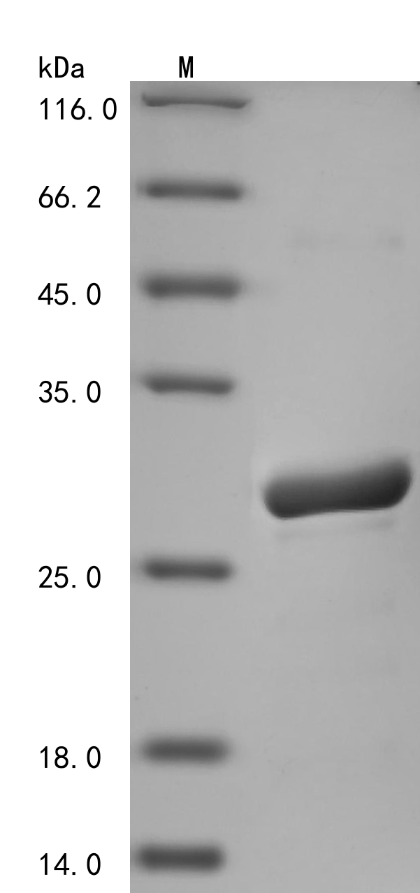
(Tris-Glycine gel) Discontinuous SDS-PAGE (reduced) with 5% enrichment gel and 15% separation gel.
DHFR Recombinant Monoclonal Antibody (CSB-RA264582A0HU)
Validated Data
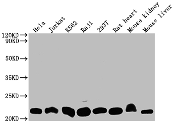
Western Blot
Positive WB detected in: Hela whole cell lysate, Jurkat whole cell lysate, K562 whole cell lysate, Raji whole cell lysate, 293T whole cell lysate, Rat heart tissue, Mouse kidney tissue, Mouse liver tissue
All lanes: DHFR antibody at 1:2000
Secondary
Goat polyclonal to rabbit IgG at 1/50000 dilution
Predicted band size: 22, 16 kDa
Observed band size: 22 kDa
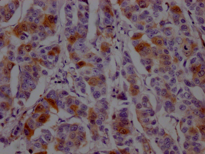
IHC image of CSB-RA264582A0HU diluted at 1:100 and staining in paraffin-embedded human breast cancer performed on a Leica BondTM system. After dewaxing and hydration, antigen retrieval was mediated by high pressure in a citrate buffer (pH 6.0). Section was blocked with 10% normal goat serum 30min at RT. Then primary antibody (1% BSA) was incubated at 4℃ overnight. The primary is detected by a Goat anti-rabbit IgG polymer labeled by HRP and visualized using 0.05% DAB.
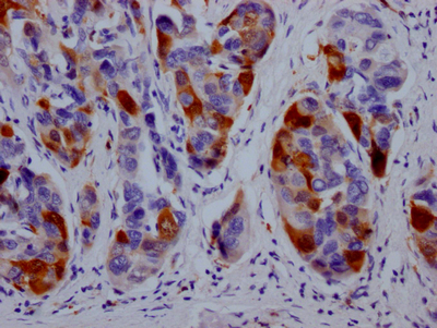
IHC image of CSB-RA264582A0HU diluted at 1:100 and staining in paraffin-embedded human liver cancer performed on a Leica BondTM system. After dewaxing and hydration, antigen retrieval was mediated by high pressure in a citrate buffer (pH 6.0). Section was blocked with 10% normal goat serum 30min at RT. Then primary antibody (1% BSA) was incubated at 4℃ overnight. The primary is detected by a Goat anti-rabbit IgG polymer labeled by HRP and visualized using 0.05% DAB.
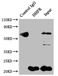
Immunoprecipitating DHFR in Hela whole cell lysate
Lane 1: Rabbit control IgG instead of CSB-RA264582A0HU in Hela whole cell lysate.
For western blotting,a HRP-conjugated Protein G antibody was used as the secondary antibody (1/2000)
Lane 2: CSB-RA264582A0HU(2µg)+ Hela whole cell lysate(500µg)
Lane 3: Hela whole cell lysate (10µg)
The following DHFR reagents supplied by CUSABIO are manufactured under a strict quality control system. Multiple applications have been validated and solid technical support is offered.
DHFR Antibodies for Homo sapiens (Human)
| Code | Product Name | Species Reactivity | Application |
|---|---|---|---|
| CSB-PA006847GA01HU | DHFR Antibody | Human,Mouse,Rat | ELISA,WB |
| CSB-PA006847HA01HU | DHFR Antibody | Human | ELISA, IHC, IF |
| CSB-RA264582A0HU | DHFR Recombinant Monoclonal Antibody | Human, Mouse, Rat | ELISA, WB, IHC, IP |
DHFR Proteins for Homo sapiens (Human)
| Code | Product Name | Source |
|---|---|---|
| CSB-YP006847HU CSB-BP006847HU CSB-MP006847HU CSB-EP006847HU-B |
Recombinant Human Dihydrofolate reductase (DHFR) | Yeast Baculovirus Mammalian cell In Vivo Biotinylation in E.coli |
| CSB-EP006847HU1 | Recombinant Human Dihydrofolate reductase (DHFR), partial | E.coli |
| CSB-EP006847HU | Recombinant Human Dihydrofolate reductase (DHFR) | E.coli |

