Call us
301-363-4651 (Available 9 a.m. to 5 p.m. CST from Monday to Friday)
MX1, also known as Myxovirus resistance 1, is an interferon - induced GTPase. It is commonly referred to as MxA. MX1 plays a crucial role in the innate immune response. It inhibits the replication of various viruses by interfering with their early stages of replication, such as uncoating and nuclear import. It is involved in the type - I interferon signaling pathway.
Dysregulation of MX1 is linked to several diseases, including viral infections like influenza and hepatitis. In the realm of drug research, MX1 has emerged as a potential therapeutic target. Scientists are exploring strategies to enhance its antiviral function, aiming to develop novel antiviral drugs to combat virus - related diseases.
MX1 Antibody (CSB-PA015249LA01HU)
Validated Data
Western Blot
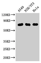
Positive WB detected in: A549 whole cell lysate, NIH/3T3 whole cell lysate, Hela whole cell lysate
All lanes: MX1 antibody at 3μg/ml
Secondary
Goat polyclonal to rabbit IgG at 1/50000 dilution
Predicted band size: 76, 56 kDa
Observed band size: 76 kDa
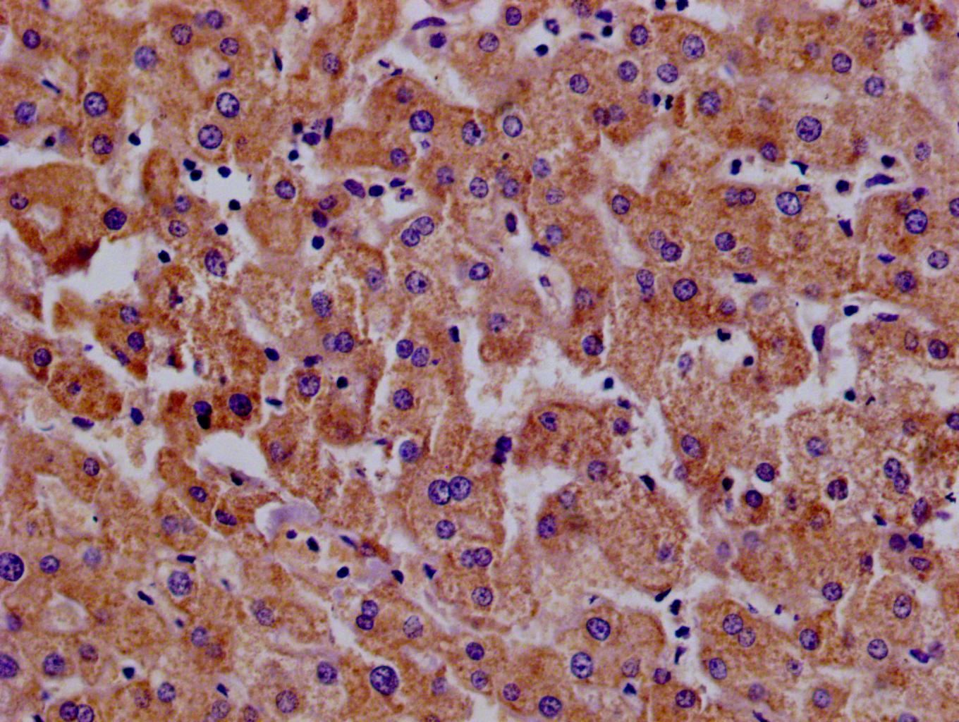
IHC image of CSB-PA015249LA01HU diluted at 1:100 and staining in paraffin-embedded human tonsil tissue performed on a Leica BondTM system. After dewaxing and hydration, antigen retrieval was mediated by high pressure in a citrate buffer (pH 6.0). Section was blocked with 10% normal goat serum 30min at RT. Then primary antibody (1% BSA) was incubated at 4°C overnight. The primary is detected by a Goat anti-rabbit polymer IgG labeled by HRP and visualized using 0.05% DAB. Secondary antibody only control: uses 1% BSA instead of primary antibody
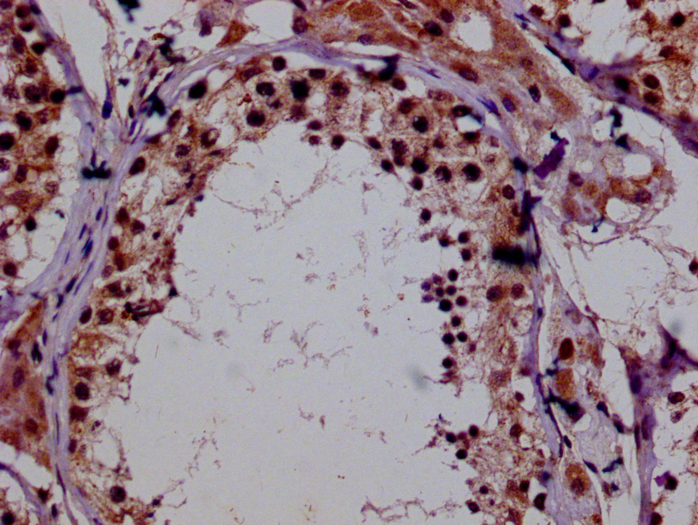
IHC image of CSB-PA015249LA01HU diluted at 1:100 and staining in paraffin-embedded human lung cancer performed on a Leica BondTM system. After dewaxing and hydration, antigen retrieval was mediated by high pressure in a citrate buffer (pH 6.0). Section was blocked with 10% normal goat serum 30min at RT. Then primary antibody (1% BSA) was incubated at 4°C overnight. The primary is detected by a Goat anti-rabbit polymer IgG labeled by HRP and visualized using 0.05% DAB. Secondary antibody only control: uses 1% BSA instead of primary antibody
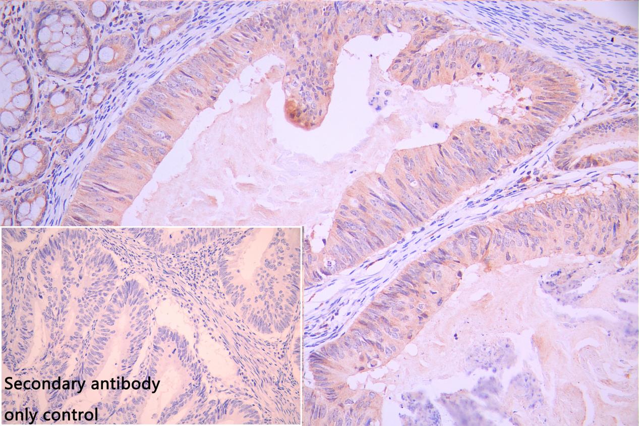
IHC image of CSB-PA015249LA01HU diluted at 1:100 and staining in paraffin-embedded human colorectal cancer performed on a Leica BondTM system. After dewaxing and hydration, antigen retrieval was mediated by high pressure in a citrate buffer (pH 6.0). Section was blocked with 10% normal goat serum 30min at RT. Then primary antibody (1% BSA) was incubated at 4°C overnight. The primary is detected by a Goat anti-rabbit polymer IgG labeled by HRP and visualized using 0.05% DAB. Secondary antibody only control: uses 1% BSA instead of primary antibody
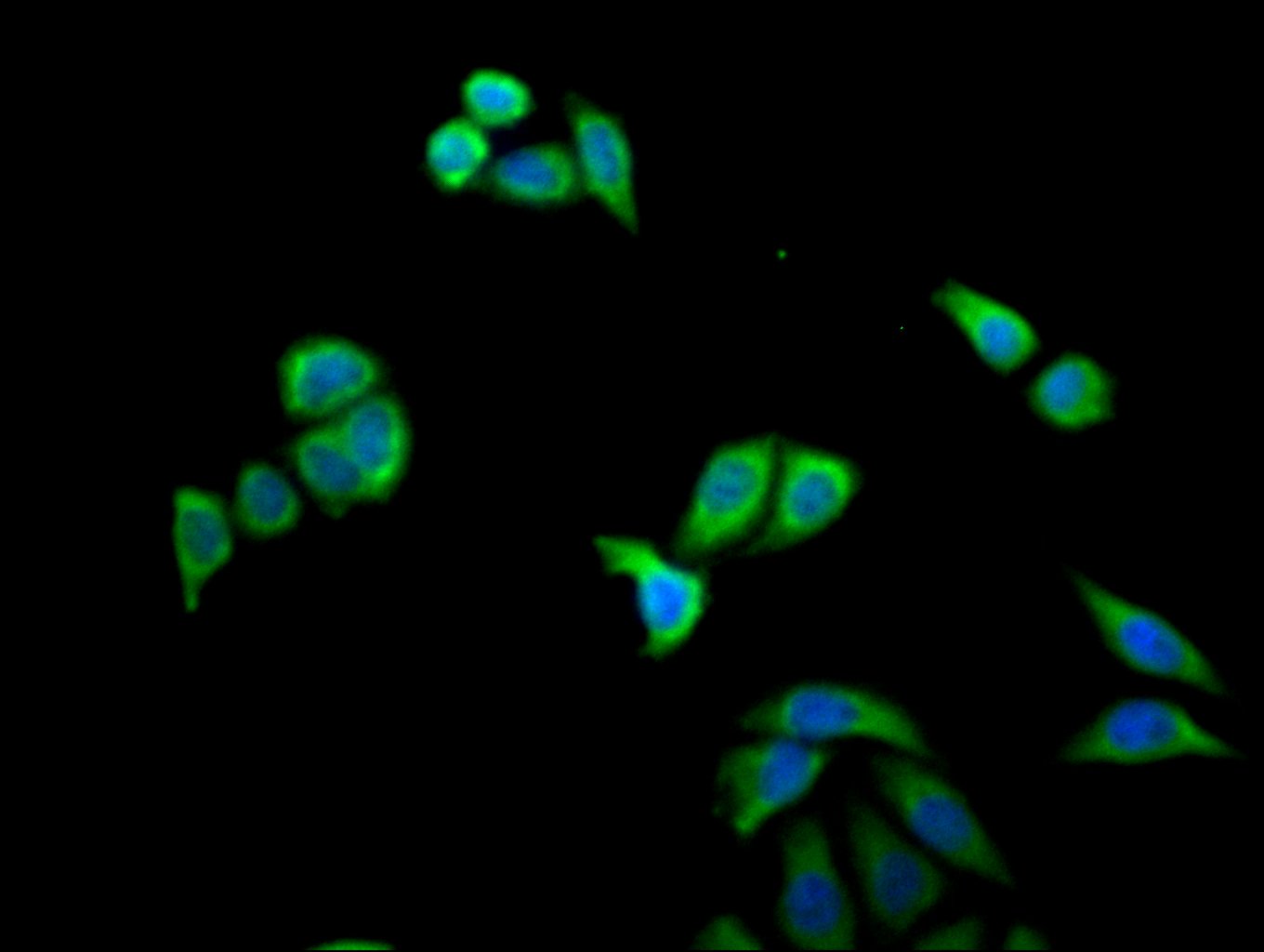
Immunofluorescence staining of PC-3 cell with CSB-PA015249LA01HU at 1:30, counter-stained with DAPI. The cells were fixed in 4% formaldehyde and and permeated by 0.2% TritonX-100 for 15 min. Then 10% normal goat serum to block non-specific protein-protein interactions . The cells were then incubated with the antibody overnight at 4℃. The secondary antibody was Alexa Fluor 488-congugated AffiniPure Goat Anti-Rabbit IgG(H+L).
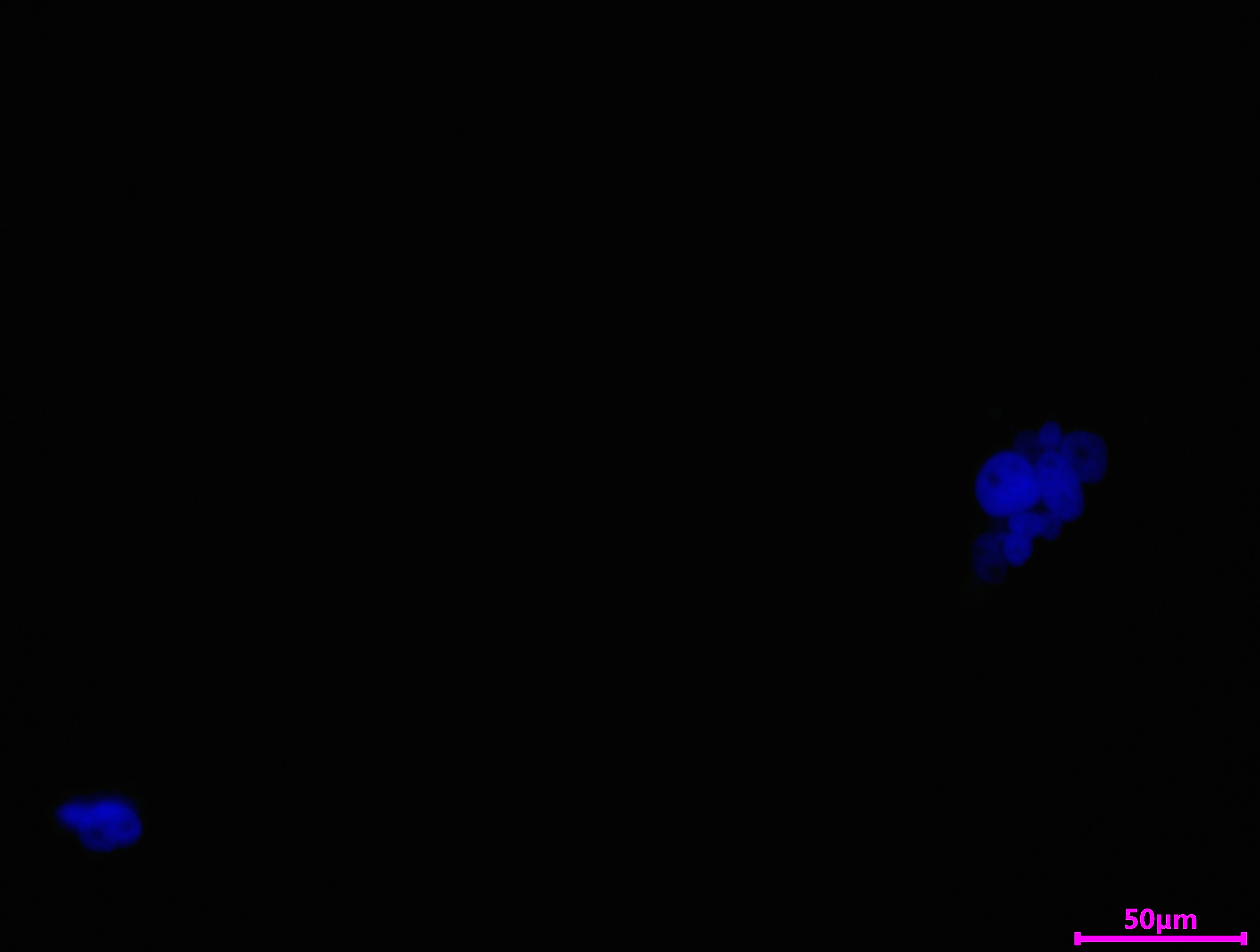
Immunofluorescence staining of PC-3 cell with 5% goat serum, counter-stained with DAPI. The cells were fixed in 4% formaldehyde and blocked in 10% normal Goat Serum. The cells were then incubated with the antibody overnight at 4C. The secondary antibody was Alexa Fluor 488-congugated AffiniPure Goat Anti-Rabbit IgG(H+L).
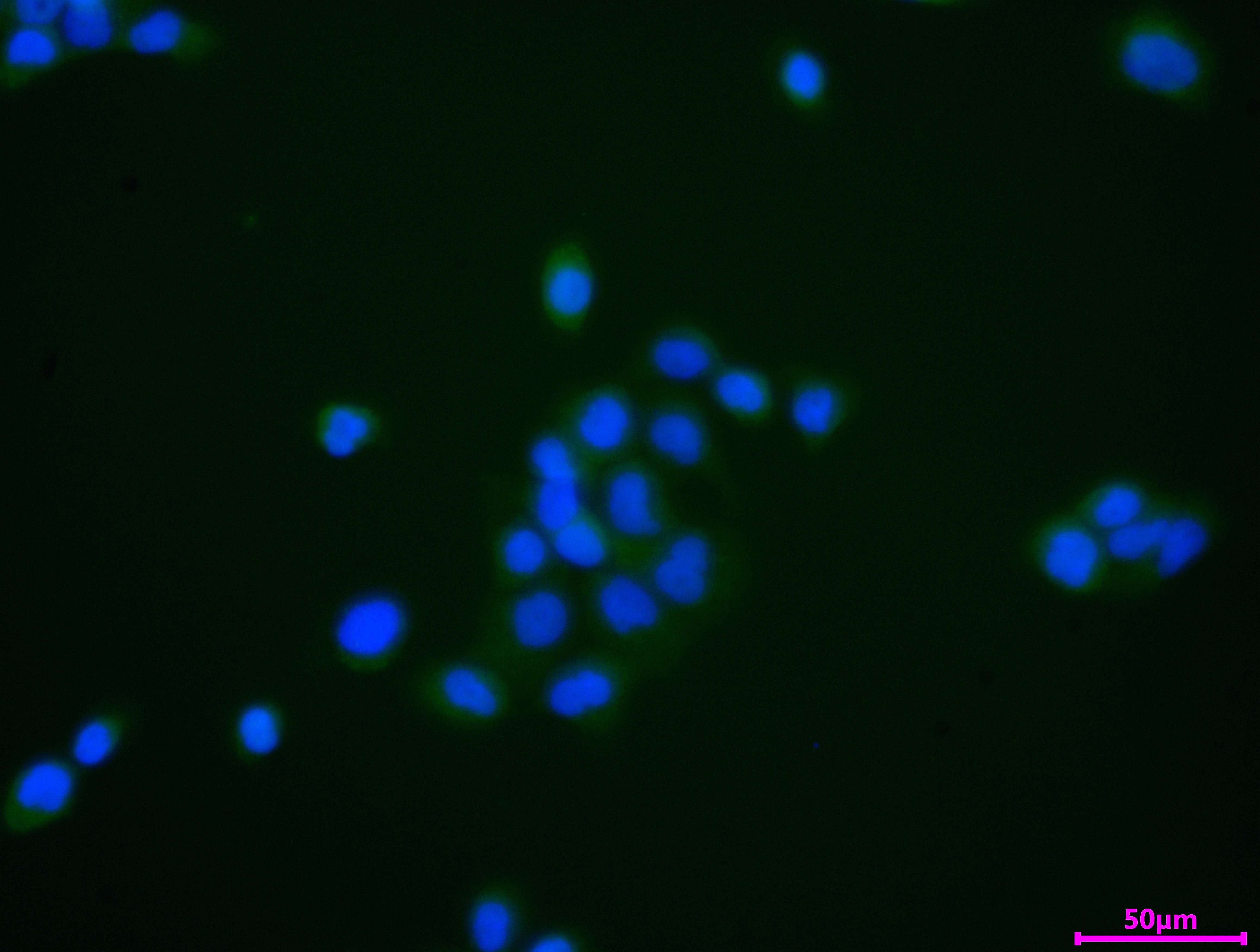
Immunofluorescence staining of A431 cell with CSB-PA015249LA01HU at 1:30, counter-stained with DAPI. The cells were fixed in 4% formaldehyde and and permeated by 0.2% TritonX-100 for 15 min. Then 10% normal goat serum to block non-specific protein-protein interactions . The cells were then incubated with the antibody overnight at 4℃. The secondary antibody was Alexa Fluor 488-congugated AffiniPure Goat Anti-Rabbit IgG(H+L).
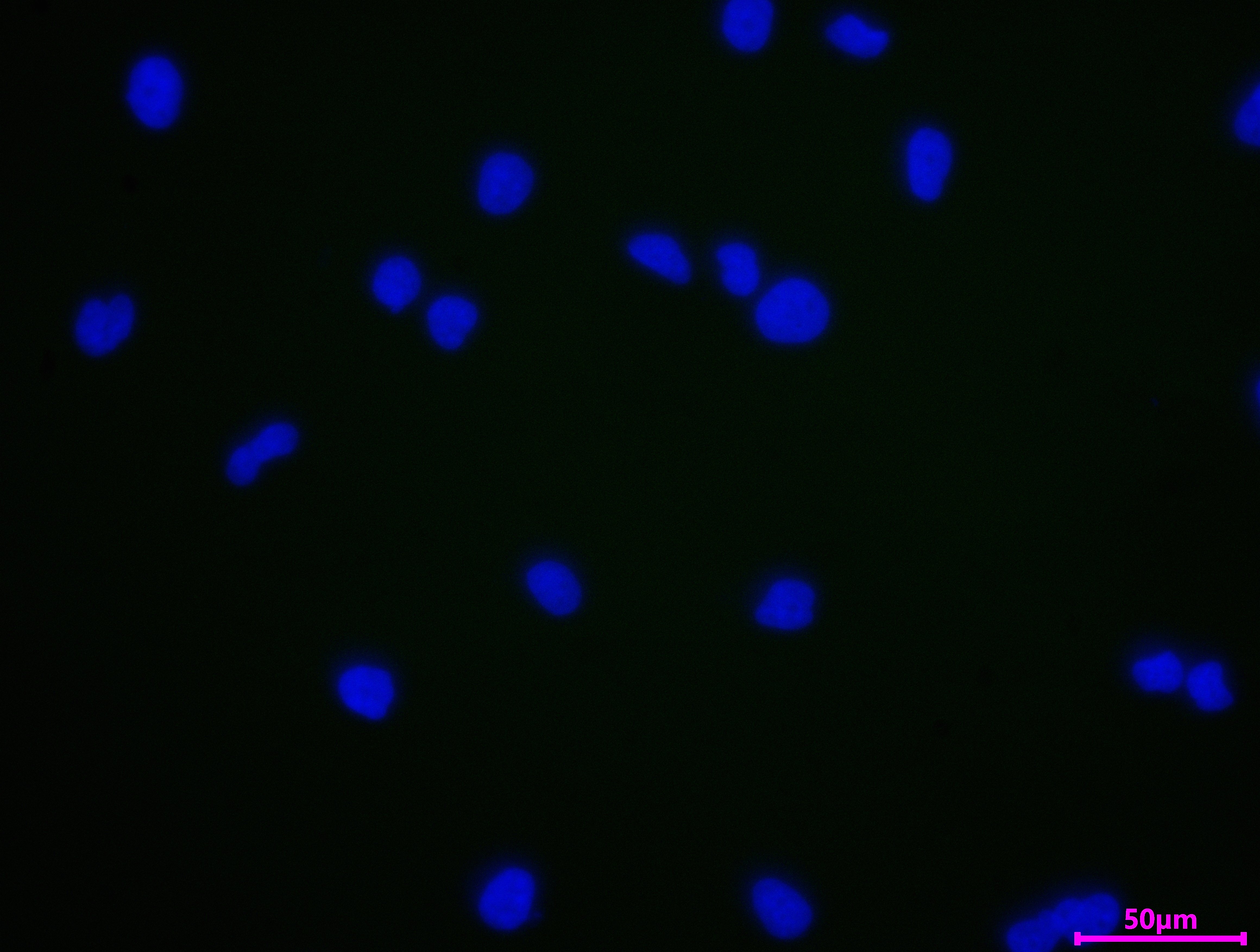
Immunofluorescence staining of A431 cell with 5% goat serum, counter-stained with DAPI. The cells were fixed in 4% formaldehyde and blocked in 10% normal Goat Serum. The cells were then incubated with the antibody overnight at 4C. The secondary antibody was Alexa Fluor 488-congugated AffiniPure Goat Anti-Rabbit IgG(H+L).
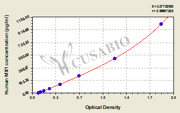
Human Interferon-induced GTP-binding protein Mx1(MX1) ELISA kit (CSB-EL015249HU)
Validated Data
Code: CSB-EL015249HU
Size: 96T,5×96T,10×96T
Sensitivity: 3.9 pg/mL
Detection Range: 15.6 pg/mL-1000 pg/mL
These standard curves are provided for demonstration only. A standard curve should be generated for each set of samples assayed.
The following MX1 reagents supplied by CUSABIO are manufactured under a strict quality control system. Multiple applications have been validated and solid technical support is offered.
MX1 Antibodies for Homo sapiens (Human)
| Code | Product Name | Species Reactivity | Application |
|---|---|---|---|
| CSB-PA015249GA01HU | MX1 Antibody | Human,Mouse | ELISA,WB,IHC |
| CSB-PA616832 | MX1 Antibody | Human | ELISA,WB |
| CSB-PA015249LA01HU | MX1 Antibody | Human, Mouse | ELISA, WB, IHC, IF |
| CSB-PA015249LB01HU | MX1 Antibody, HRP conjugated | Human | ELISA |
| CSB-PA015249LC01HU | MX1 Antibody, FITC conjugated | Human | |
| CSB-PA015249LD01HU | MX1 Antibody, Biotin conjugated | Human | ELISA |
MX1 Proteins for Homo sapiens (Human)
| Code | Product Name | Source |
|---|---|---|
| CSB-YP015249HU CSB-EP015249HU CSB-BP015249HU CSB-MP015249HU CSB-EP015249HU-B |
Recombinant Human Interferon-induced GTP-binding protein Mx1 (MX1) | Yeast E.coli Baculovirus Mammalian cell In Vivo Biotinylation in E.coli |
MX1 ELISA Kit for Homo sapiens (Human)
| Code | Product Name | Sample Types | Sensitivity |
|---|---|---|---|
| CSB-EL015249HU | Human Interferon-induced GTP-binding protein Mx1(MX1) ELISA kit | serum, plasma, urine, tissue homogenates, cell lysates | 3.9 pg/mL |

