Call us
301-363-4651 (Available 9 a.m. to 5 p.m. CST from Monday to Friday)
PRKCH encodes Protein Kinase C Eta (PKCη), a novel serine/threonine kinase of the PKC family. Activated by diacylglycerol (DAG) independent of calcium, it mediates signal transduction pathways regulating cell proliferation, differentiation, and stress responses, with high expression in epithelial tissues such as keratinocytes.
Genetic variants of PRKCH are linked to increased ischemic stroke risk in East Asian populations, and its dysregulation contributes to skin disorders like psoriasis. Ongoing drug development includes small-molecule inhibitors targeting PKCη, aiming to modulate its activity for stroke prevention and psoriasis treatment.
Recombinant Human Protein kinase C eta type (PRKCH) (CSB-EP018706HU)
Validated Data
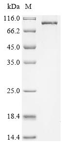
(Tris-Glycine gel) Discontinuous SDS-PAGE (reduced) with 5% enrichment gel and 15% separation gel.
PRKCH Antibody (CSB-PA15879A0Rb)
Validated Data
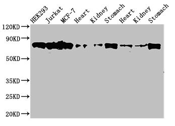
Western Blot
Positive WB detected in: HEK293 whole cell lysate, Jurkat whole cell lysate, MCF-7 whole cell lysate, Rat heart tissue, Rat kideny tissue, Rat stomach tissue, Mouse heart tissue, Mouse kidney tissue, Mouse stomach tissue
All lanes: PRKCH antibody at 3.5µg/ml
Secondary
Goat polyclonal to rabbit IgG at 1/50000 dilution
Predicted band size: 78, 60 kDa
Observed band size: 78 kDa
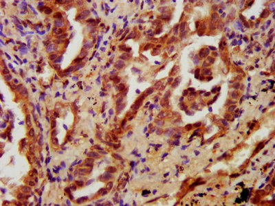
IHC image of CSB-PA15879A0Rb diluted at 1:700 and staining in paraffin-embedded human lung cancer performed on a Leica BondTM system. After dewaxing and hydration, antigen retrieval was mediated by high pressure in a citrate buffer (pH 6.0). Section was blocked with 10% normal goat serum 30min at RT. Then primary antibody (1% BSA) was incubated at 4°C overnight. The primary is detected by a biotinylated secondary antibody and visualized using an HRP conjugated SP system.
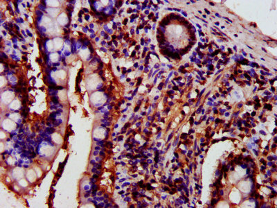
IHC image of CSB-PA15879A0Rb diluted at 1:700 and staining in paraffin-embedded human small intestine tissue performed on a Leica BondTM system. After dewaxing and hydration, antigen retrieval was mediated by high pressure in a citrate buffer (pH 6.0). Section was blocked with 10% normal goat serum 30min at RT. Then primary antibody (1% BSA) was incubated at 4°C overnight. The primary is detected by a biotinylated secondary antibody and visualized using an HRP conjugated SP system.
The following PRKCH reagents supplied by CUSABIO are manufactured under a strict quality control system. Multiple applications have been validated and solid technical support is offered.
PRKCH Antibodies for Homo sapiens (Human)
| Code | Product Name | Species Reactivity | Application |
|---|---|---|---|
| CSB-PA15879A0Rb | PRKCH Antibody | Human, Mouse, Rat | ELISA, WB, IHC |
| CSB-PA15879B0Rb | PRKCH Antibody, HRP conjugated | Human | ELISA |
| CSB-PA15879C0Rb | PRKCH Antibody, FITC conjugated | Human | |
| CSB-PA15879D0Rb | PRKCH Antibody, Biotin conjugated | Human | ELISA |
PRKCH Proteins for Homo sapiens (Human)
| Code | Product Name | Source |
|---|---|---|
| CSB-YP018706HU CSB-BP018706HU CSB-MP018706HU CSB-EP018706HU-B |
Recombinant Human Protein kinase C eta type (PRKCH) | Yeast Baculovirus Mammalian cell In Vivo Biotinylation in E.coli |
| CSB-EP018706HU | Recombinant Human Protein kinase C eta type (PRKCH) | E.coli |
| CSB-EP018706HUa0 | Recombinant Human Protein kinase C eta type (PRKCH) | E.coli |
PRKCH Proteins for Mus musculus (Mouse)
| Code | Product Name | Source |
|---|---|---|
| CSB-YP018706MO CSB-EP018706MO CSB-BP018706MO CSB-MP018706MO CSB-EP018706MO-B |
Recombinant Mouse Protein kinase C eta type (Prkch) | Yeast E.coli Baculovirus Mammalian cell In Vivo Biotinylation in E.coli |
PRKCH Proteins for Rattus norvegicus (Rat)
| Code | Product Name | Source |
|---|---|---|
| CSB-YP723752RA CSB-EP723752RA CSB-BP723752RA CSB-MP723752RA CSB-EP723752RA-B |
Recombinant Rat Protein kinase C eta type (Prkch) | Yeast E.coli Baculovirus Mammalian cell In Vivo Biotinylation in E.coli |

