Call us
301-363-4651 (Available 9 a.m. to 5 p.m. CST from Monday to Friday)
| Code | CSB-RA018699A638phHU |
| Size | US$210 |
| Order now | |
| Image |
|
| Have Questions? | Leave a Message or Start an on-line Chat |
| Application | Recommended Dilution |
|---|---|
| WB | 1:500-1:5000 |
| IF | 1:20-1:200 |
Protein kinase C alpha (PRKCA/PKC-alpha) serves as a central node in cellular signal transduction, mediating responses to growth factors, hormones, and other extracellular signals that drive proliferation, differentiation, and survival. Phosphorylation at threonine 638 represents a critical regulatory event in the enzyme's catalytic maturation, making this modification an important readout for researchers investigating PKC-dependent signaling cascades in cancer biology, metabolic regulation, and immune cell function.
This recombinant monoclonal antibody, generated against a synthetic phosphopeptide corresponding to the human phospho-PRKCA T638 region, offers the reproducibility and sequence-defined specificity that demanding phosphoprotein studies require. Because recombinant production eliminates the batch variability inherent to traditional hybridoma methods, researchers can confidently compare results across extended experimental timelines and between laboratories.
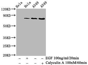
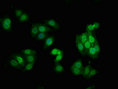
Validation studies confirm reliable performance across multiple detection platforms. In western blot applications, the antibody detects a band at the predicted 80 kDa molecular weight in HeLa whole cell lysates and in A549 cells following treatment with Calyculin A or EGF, demonstrating its utility for monitoring phosphorylation dynamics under different stimulation conditions. Recommended working dilutions range from 1:500 to 1:5000, providing flexibility to optimize signal-to-background ratios across different sample preparations. Immunofluorescence validation in HepG2 cells at 1:100 dilution reveals clear subcellular localization patterns, enabling spatial analysis of activated PKC-alpha within intact cells.
Whether investigating receptor-mediated signaling pathways, screening kinase inhibitor efficacy, or characterizing phosphorylation-dependent protein interactions, this antibody provides a dependable tool for signal transduction research focused on human samples.
There are currently no reviews for this product.

