Call us
301-363-4651 (Available 9 a.m. to 5 p.m. CST from Monday to Friday)
APMAP (Adipocyte Plasma Membrane Associated Protein), also known as adipocyte membrane protein, is a transmembrane protein highly expressed in adipocytes. It regulates adipocyte differentiation and lipid homeostasis by modulating insulin signaling pathways, facilitating glucose uptake and triglyceride storage in adipose tissue.
Dysregulation of APMAP links to metabolic disorders like obesity and type 2 diabetes—reduced expression correlates with insulin resistance. Preclinical studies explore targeting APMAP to enhance adipocyte function; preliminary data shows modulating its levels may improve glucose tolerance, though no clinical drugs targeting it have entered trials yet.
Recombinant Human Adipocyte plasma membrane-associated protein (APMAP), partial (CSB-EP884629HUb1)
Validated Data
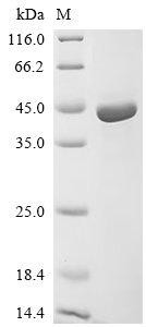
(Tris-Glycine gel) Discontinuous SDS-PAGE (reduced) with 5% enrichment gel and 15% separation gel.
APMAP Antibody (CSB-PA884629LA01HU)
Validated Data
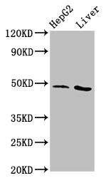
Western Blot
Positive WB detected in: HepG2 whole cell lysate, Rat liver tissue
All lanes: APMAP antibody at 3.1µg/ml
Secondary
Goat polyclonal to rabbit IgG at 1/50000 dilution
Predicted band size: 47, 33 kDa
Observed band size: 47 kDa
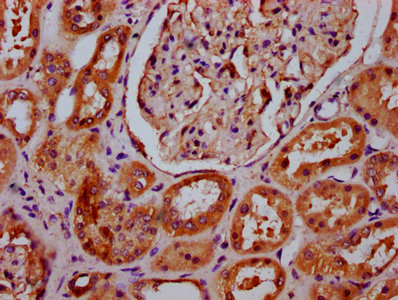
IHC image of CSB-PA884629LA01HU diluted at 1:400 and staining in paraffin-embedded human kidney tissue performed on a Leica BondTM system. After dewaxing and hydration, antigen retrieval was mediated by high pressure in a citrate buffer (pH 6.0). Section was blocked with 10% normal goat serum 30min at RT. Then primary antibody (1% BSA) was incubated at 4°C overnight. The primary is detected by a biotinylated secondary antibody and visualized using an HRP conjugated SP system.
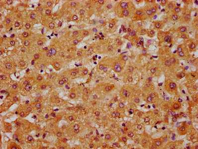
IHC image of CSB-PA884629LA01HU diluted at 1:400 and staining in paraffin-embedded human liver tissue performed on a Leica BondTM system. After dewaxing and hydration, antigen retrieval was mediated by high pressure in a citrate buffer (pH 6.0). Section was blocked with 10% normal goat serum 30min at RT. Then primary antibody (1% BSA) was incubated at 4°C overnight. The primary is detected by a biotinylated secondary antibody and visualized using an HRP conjugated SP system.
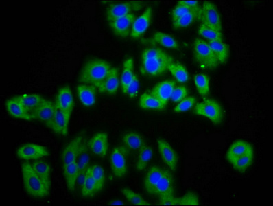
Immunofluorescence staining of HepG2 cells with CSB-PA884629LA01HU at 1:133, counter-stained with DAPI. The cells were fixed in 4% formaldehyde, permeabilized using 0.2% Triton X-100 and blocked in 10% normal Goat Serum. The cells were then incubated with the antibody overnight at 4°C. The secondary antibody was Alexa Fluor 488-congugated AffiniPure Goat Anti-Rabbit IgG(H+L).
The following APMAP reagents supplied by CUSABIO are manufactured under a strict quality control system. Multiple applications have been validated and solid technical support is offered.
APMAP Antibodies for Homo sapiens (Human)
| Code | Product Name | Species Reactivity | Application |
|---|---|---|---|
| CSB-PA165039 | APMAP Antibody | Human | ELISA,WB,IHC |
| CSB-PA915948 | APMAP Antibody | Human | ELISA,WB,IHC |
| CSB-PA884629LA01HU | APMAP Antibody | Human, Rat | ELISA, WB, IHC, IF |
APMAP Proteins for Homo sapiens (Human)
| Code | Product Name | Source |
|---|---|---|
| CSB-YP884629HU CSB-EP884629HU CSB-BP884629HU CSB-EP884629HU-B |
Recombinant Human Adipocyte plasma membrane-associated protein (APMAP), partial | Yeast E.coli Baculovirus In Vivo Biotinylation in E.coli |
| CSB-EP884629HUb1 | Recombinant Human Adipocyte plasma membrane-associated protein (APMAP), partial | E.coli |
| CSB-MP884629HU | Recombinant Human Adipocyte plasma membrane-associated protein (APMAP), partial | Mammalian cell |
| CSB-MP884629HUd7 | Recombinant Human Adipocyte plasma membrane-associated protein (APMAP), partial | Mammalian cell |

