Call us
301-363-4651 (Available 9 a.m. to 5 p.m. CST from Monday to Friday)
FUBP1 encodes far upstream element-binding protein 1, a DNA/RNA-binding protein also known as FBP or FUBP. It regulates transcription by binding to far upstream elements in gene promoters, particularly activating c-MYC expression. FUBP1 participates in cell cycle progression and proliferation signaling pathways, including the Wnt/β-catenin and MAPK pathways.
Aberrant FUBP1 expression is linked to several cancers, such as hepatocellular carcinoma and colorectal cancer, where it promotes tumor growth and metastasis. Preclinical studies investigate small molecule inhibitors targeting FUBP1-DNA interactions to disrupt c-MYC-driven oncogenesis, though no clinical candidates have advanced to trials yet.
FUBP1 Recombinant Monoclonal Antibody (CSB-RA157765A0HU)
Validated Data
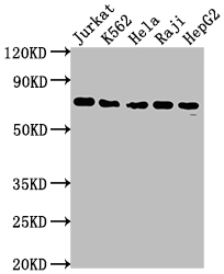
Western Blot
Positive WB detected in: Jurkat whole cell lysate, K562 whole cell lysate, Hela whole cell lysate, Raji whole cell lysate, HepG2 whole cell lysate
All lanes: FUBP1 antibody at 1:2000
Secondary
Goat polyclonal to rabbit IgG at 1/50000 dilution
Predicted band size: 68, 69 kDa
Observed band size: 69 kDa
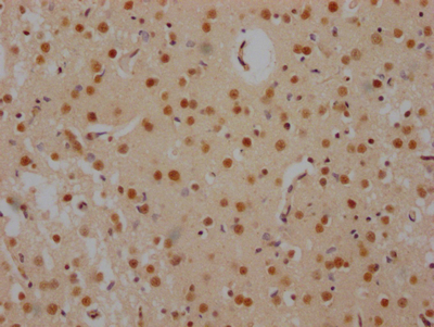
IHC image of CSB-RA157765A0HU diluted at 1:100 and staining in paraffin-embedded human brain tissue performed on a Leica BondTM system. After dewaxing and hydration, antigen retrieval was mediated by high pressure in a citrate buffer (pH 6.0). Section was blocked with 10% normal goat serum 30min at RT. Then primary antibody (1% BSA) was incubated at 4℃ overnight. The primary is detected by a Goat anti-rabbit IgG polymer labeled by HRP and visualized using 0.05% DAB.
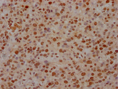
IHC image of CSB-RA157765A0HU diluted at 1:100 and staining in paraffin-embedded human glioma cancer performed on a Leica BondTM system. After dewaxing and hydration, antigen retrieval was mediated by high pressure in a citrate buffer (pH 6.0). Section was blocked with 10% normal goat serum 30min at RT. Then primary antibody (1% BSA) was incubated at 4℃ overnight. The primary is detected by a Goat anti-rabbit IgG polymer labeled by HRP and visualized using 0.05% DAB.
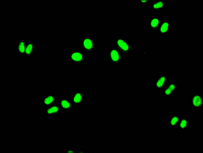
Immunofluorescence staining of Hela Cells with CSB-RA157765A0HU at 1:50, counter-stained with DAPI. The cells were fixed in 4% formaldehyde, permeated by 0.2% TritonX-100, and blocked in 10% normal Goat Serum. The cells were then incubated with the antibody overnight at 4℃. Nuclear DNA was labeled in blue with DAPI. The secondary antibody was FITC-conjugated AffiniPure Goat Anti-Rabbit IgG (H+L).
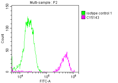
Overlay histogram showing Jurkat cells stained with CSB-RA157765A0HU (red line) at 1:50. The cells were fixed with 70% Ethylalcohol (18h) and then incubated in 10% normal goat serum to block non-specific protein-protein interactions followedby the antibody (1µg/1*106 cells) for 1 h at 4℃.The secondary antibody used was FITC-conjugated goat anti-rabbit IgG (H+L) at 1/200 dilution for 30min at 4℃. Control antibody (green line) was Rabbit IgG (1µg/1*106 cells) used under the same conditions. Acquisition of >10,000 events was performed.
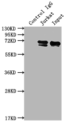
Immunoprecipitating FUBP1 in Jurkat whole cell lysate
Lane 1: Rabbit control IgG instead of CSB-RA157765A0HU in Jurkat whole cell lysate.
For western blotting,a HRP-conjugated Protein G antibody was used as the secondary antibody (1/2000)
Lane 2: CSB-RA157765A0HU(2µg)+ Jurkat whole cell lysate(500µg)
Lane 3: Jurkat whole cell lysate (10µg)
The following FUBP1 reagents supplied by CUSABIO are manufactured under a strict quality control system. Multiple applications have been validated and solid technical support is offered.
FUBP1 Antibodies for Homo sapiens (Human)
| Code | Product Name | Species Reactivity | Application |
|---|---|---|---|
| CSB-RA157765A0HU | FUBP1 Recombinant Monoclonal Antibody | Human | ELISA, WB, IHC, IF, FC, IP |
FUBP1 Proteins for Homo sapiens (Human)
| Code | Product Name | Source |
|---|---|---|
| CSB-YP846584HU CSB-EP846584HU CSB-BP846584HU CSB-MP846584HU CSB-EP846584HU-B |
Recombinant Human Far upstream element-binding protein 1 (FUBP1) | Yeast E.coli Baculovirus Mammalian cell In Vivo Biotinylation in E.coli |

