Call us
301-363-4651 (Available 9 a.m. to 5 p.m. CST from Monday to Friday)
The FUS gene encodes the Fused in Sarcoma (FUS) protein, also known as Translocated in Liposarcoma (TLS). As an RNA-binding protein, FUS regulates pre-mRNA splicing, mRNA transport, and translational processes, interacting with core RNA processing machinery to maintain cellular RNA homeostasis. It integrates into pathways governing gene expression and stress response.
Mutations in FUS are associated with amyotrophic lateral sclerosis (ALS), frontotemporal dementia (FTD), and liposarcoma. For ALS/FTD, drug development targets FUS aggregation— a key pathogenic event— using small molecules that inhibit misfolding or RNA interference to reduce toxic protein levels, though most candidates are in preclinical or early clinical phases.
Recombinant Human RNA-binding protein FUS (FUS) (CSB-EP009069HU)
Validated Data
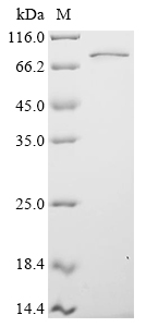
(Tris-Glycine gel) Discontinuous SDS-PAGE (reduced) with 5% enrichment gel and 15% separation gel.
FUS Recombinant Monoclonal Antibody (CSB-RA796544A0HU)
Validated Data
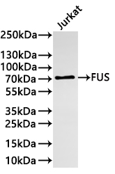
Western Blot
Positive WB detected in:Jurkat whole cell lysate(30µg)
All lanes: FUS antibody at 1:1000
Secondary
Goat polyclonal to rabbit IgG at 1/40000 dilution
Predicted band size: 53 kDa
Observed band size: 70 kDa
Exposure time:2min
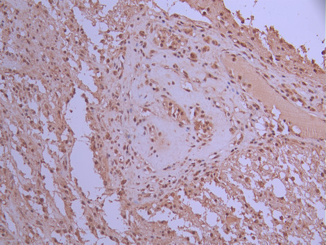
IHC image of CSB-RA796544A0HU diluted at 1:100 and staining in paraffin-embedded human glioma cancer performed on a Leica BondTM system. After dewaxing and hydration, antigen retrieval was mediated by high pressure in a citrate buffer (pH 6.0). Section was blocked with 10% normal goat serum 30min at RT. Then primary antibody (1% BSA) was incubated at 4°C overnight. The primary is detected by a Goat anti-rabbit polymer IgG labeled by HRP and visualized using 0.05% DAB.
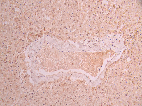
IHC image of CSB-RA796544A0HU diluted at 1:100 and staining in paraffin-embedded human liver tissue performed on a Leica BondTM system. After dewaxing and hydration, antigen retrieval was mediated by high pressure in a citrate buffer (pH 6.0). Section was blocked with 10% normal goat serum 30min at RT. Then primary antibody (1% BSA) was incubated at 4°C overnight. The primary is detected by a Goat anti-rabbit polymer IgG labeled by HRP and visualized using 0.05% DAB.
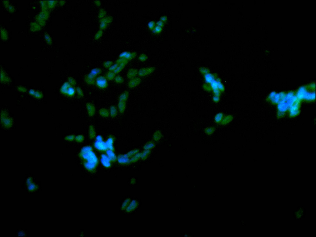
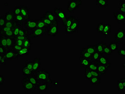
Immunofluorescence staining of SH-SY5Y cell with CSB-RA796544A0HU at 1:50, counter-stained with DAPI. The cells were fixed in 4% formaldehyde, permeabilized using 0.2% Triton X-100 and blocked in 10% normal Goat Serum. The cells were then incubated with the antibody overnight at 4°C. The secondary antibody was Alexa Fluor 488-congugated AffiniPure Goat Anti-Rabbit IgG(H+L).
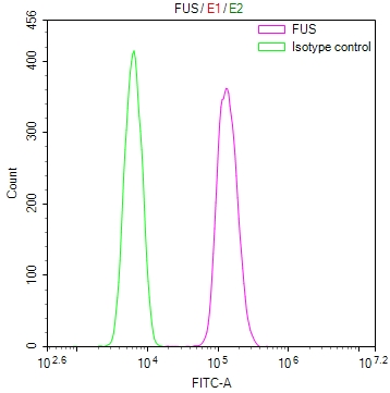
Overlay Peak curve showing SH-SY5Y cells stained with CSB-RA796544A0HU (red line) at 1:100. The cells were fixed in 4% formaldehyde and permeated by 0.2% TritonX-100 for 10min. Then 10% normal goat serum to block non-specific protein-protein interactions followed by the antibody (1ug/1*106cells) for 45min at 4℃. The secondary antibody used was FITC-conjugated goat anti-rabbit IgG (H+L) at 1/200 dilution for 35min at 4℃.Control antibody (green line) was Rabbit IgG (1ug/1*106cells) used under the same conditions. Acquisition of >10,000 events was performed.
FUS Antibody (CSB-PA02704A0Rb)
Validated Data
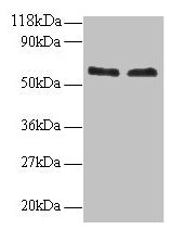
Western blot
All lanes: FUS antibody at 2µg/ml
Lane 1: 293T whole cell lysate
Lane 2: EC109 whole cell lysate
Secondary
Goat polyclonal to rabbit IgG at 1/15000 dilution
Predicted band size: 54 kDa
Observed band size: 60 kDa
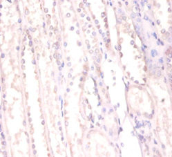
Immunohistochemistry of paraffin-embedded human kidney tissue using CSB-PA02704A0Rb at dilution of 1:100
Immunofluorescent analysis of HepG2 cells using CSB-PA02704A0Rb at dilution of 1:100 and Alexa Fluor 488-congugated AffiniPure Goat Anti-Rabbit IgG(H+L)
Human RNA-binding protein FUS (FUS/TLS) ELISA kit (CSB-E17376h)
Validated Data
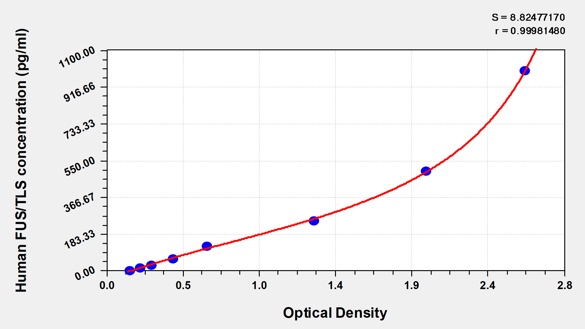
Code: CSB-E17376h
Size: 96T,5×96T,10×96T
Sensitivity: 3.9 pg/mL
Detection Range: 15.6 pg/mL-1000 pg/mL
These standard curves are provided for demonstration only. A standard curve should be generated for each set of samples assayed.
The following FUS reagents supplied by CUSABIO are manufactured under a strict quality control system. Multiple applications have been validated and solid technical support is offered.
FUS Antibodies for Homo sapiens (Human)
| Code | Product Name | Species Reactivity | Application |
|---|---|---|---|
| CSB-PA02704A0Rb | FUS Antibody | Human | ELISA, WB, IHC, IF |
| CSB-PA02704D0Rb | FUS Antibody, Biotin conjugated | Human | ELISA |
| CSB-PA009069GA01HU | FUS Antibody | Human,Mouse,Rat | ELISA,WB,IHC,IF |
| CSB-PA229936 | FUS Antibody | Human,Mouse | ELISA,IHC |
| CSB-PA815873 | FUS Antibody | Human,Mouse | ELISA,IHC |
| CSB-RA796544A0HU | FUS Recombinant Monoclonal Antibody | Human | ELISA, WB, IHC, IF, FC |
FUS Proteins for Homo sapiens (Human)
| Code | Product Name | Source |
|---|---|---|
| CSB-YP009069HU CSB-BP009069HU CSB-MP009069HU CSB-EP009069HU-B |
Recombinant Human RNA-binding protein FUS (FUS) | Yeast Baculovirus Mammalian cell In Vivo Biotinylation in E.coli |
| CSB-EP009069HU | Recombinant Human RNA-binding protein FUS (FUS) | E.coli |
| CSB-EP009069HUe7 | Recombinant Human RNA-binding protein FUS (FUS) | E.coli |
| CSB-EP009069HUc7 | Recombinant Human RNA-binding protein FUS (FUS) | E.coli |
FUS ELISA Kit for Homo sapiens (Human)
| Code | Product Name | Sample Types | Sensitivity |
|---|---|---|---|
| CSB-E17376h | Human RNA-binding protein FUS (FUS/TLS) ELISA kit | serum, plasma, cell lysates | 3.9 pg/mL |

