Call us
301-363-4651 (Available 9 a.m. to 5 p.m. CST from Monday to Friday)
MTAP encodes methylthioadenosine phosphorylase, an enzyme also called MTAPase. It catalyzes the breakdown of S-methyl-5'-thioadenosine (MTA) into adenine and 5-methylthio-D-ribose 1-phosphate, a key step in purine salvage metabolism. Loss of MTAP leads to MTA accumulation, activating the mTOR signaling pathway to promote cell proliferation.
MTAP deletion is frequent in glioblastoma, lung cancer, and melanoma, often co-occurring with CDKN2A loss on chromosome 9p21. Drug development targeting MTAP-deficient tumors is ongoing: mTOR inhibitors like everolimus show preclinical efficacy, while agents blocking MTA-mediated signaling are explored as potential therapies.
MTAP Recombinant Monoclonal Antibody (CSB-RA299565A0HU)
Validated Data
Western Blot
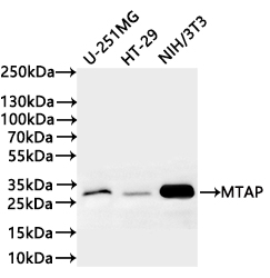
Positive WB detected in: U-251MG whole cell lysate(20µg),HT-29 whole cell lysate(20µg), NIH/3T3 whole cell lysate(20µg)
All lanes: MTAP antibody at 1:1000
Secondary
Goat polyclonal to rabbit IgG at 1/40000 dilution
Predicted band size: 31 kDa
Observed band size: 31 kDa
Exposure time:1min20s
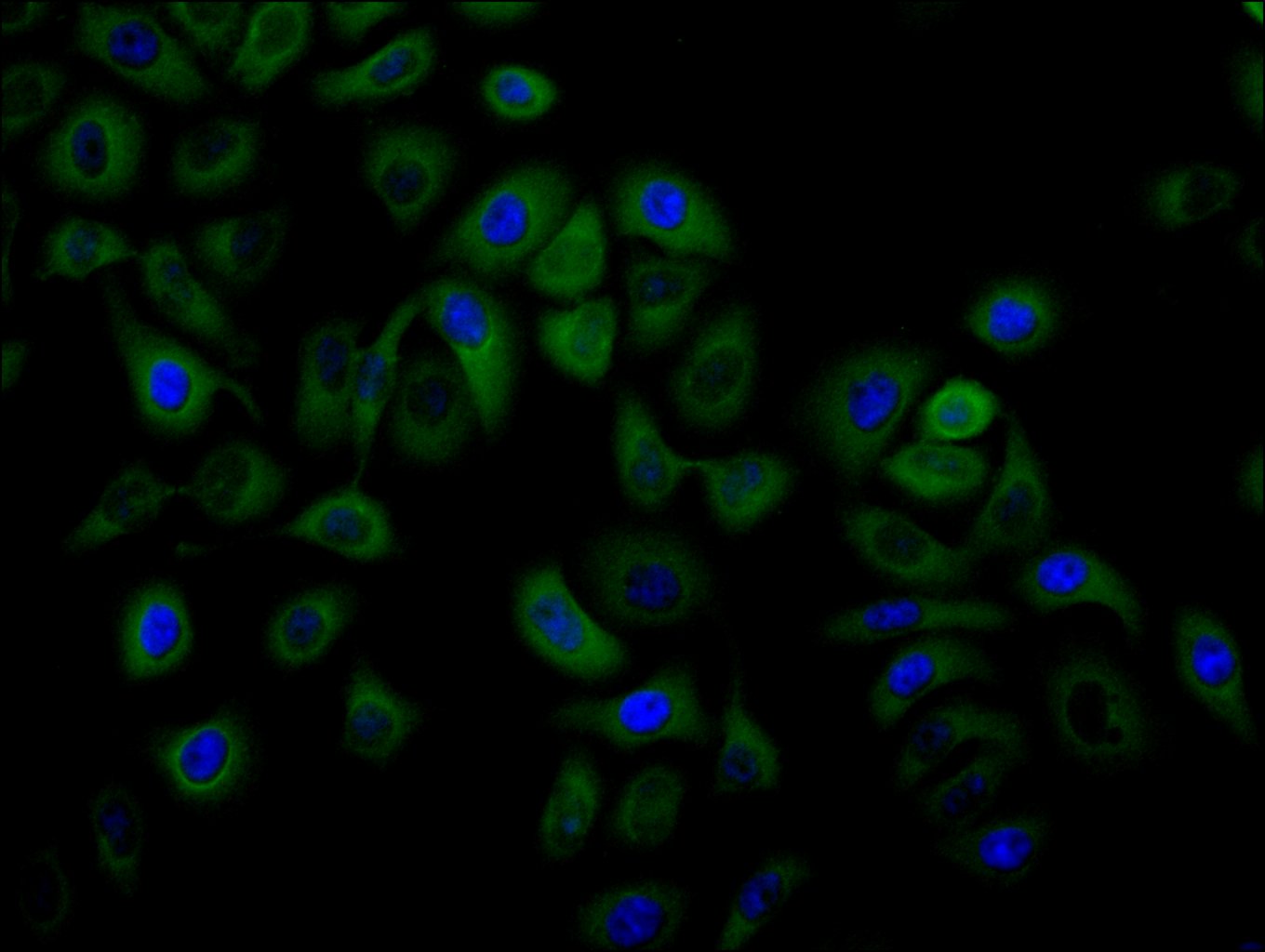
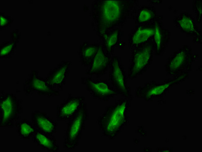
Immunofluorescence staining of Hela cell with CSB-RA299565A0HU at 1:50, counter-stained with DAPI. The cells were fixed in 4% formaldehyde, permeabilized using 0.2% Triton X-100 and blocked in 10% normal Goat Serum. The cells were then incubated with the antibody overnight at 4°C. The secondary antibody was Alexa Fluor 488-congugated AffiniPure Goat Anti-Rabbit IgG(H+L).
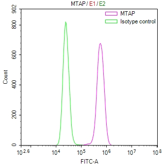
Overlay Peak curve showing Hela cells stained with CSB-RA299565A0HU (red line) at 1:100. The cells were fixed in 4% formaldehyde and permeated by 0.2% TritonX-100 for 10min. Then 10% normal goat serum to block non-specific protein-protein interactions followed by the antibody (1ug/1*106cells) for 45min at 4℃. The secondary antibody used was FITC-conjugated goat anti-rabbit IgG (H+L) at 1/200 dilution for 35min at 4℃.Control antibody (green line) was Rabbit IgG (1ug/1*106cells) used under the same conditions. Acquisition of >10,000 events was performed.
MTAP Antibody (CSB-PA622639LA01HU)
Validated Data
Western Blot
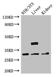
Positive WB detected in: NIH/3T3 whole cell lysate, Mouse liver tissue, Mouse kidney tissue
All lanes: MTAP antibody at 2.7μg/ml
Secondary
Goat polyclonal to rabbit IgG at 1/50000 dilution
Predicted band size: 32, 39, 37, 31, 34, 33, 27 kDa
Observed band size: 32, 39 kDa
Immunofluorescent analysis of U251 cells using CSB-PA622639LA01HU at dilution of 1:100 and Alexa Fluor 488-congugated AffiniPure Goat Anti-Rabbit IgG(H+L)
The following MTAP reagents supplied by CUSABIO are manufactured under a strict quality control system. Multiple applications have been validated and solid technical support is offered.
MTAP Antibodies for Homo sapiens (Human)
| Code | Product Name | Species Reactivity | Application |
|---|---|---|---|
| CSB-PA622639LA01HU | MTAP Antibody | Human, Mouse | ELISA, WB, IF |
| CSB-PA622639LB01HU | MTAP Antibody, HRP conjugated | Human | ELISA |
| CSB-PA622639LC01HU | MTAP Antibody, FITC conjugated | Human | |
| CSB-PA622639LD01HU | MTAP Antibody, Biotin conjugated | Human | ELISA |
| CSB-RA299565A0HU | MTAP Recombinant Monoclonal Antibody | Human, Mouse | ELISA, WB, IF, FC |
MTAP Proteins for Homo sapiens (Human)
| Code | Product Name | Source |
|---|---|---|
| CSB-YP622639HU CSB-EP622639HU CSB-BP622639HU CSB-MP622639HU CSB-EP622639HU-B |
Recombinant Human S-methyl-5'-thioadenosine phosphorylase (MTAP) | Yeast E.coli Baculovirus Mammalian cell In Vivo Biotinylation in E.coli |

