Call us
301-363-4651 (Available 9 a.m. to 5 p.m. CST from Monday to Friday)
PEBP1 encodes phosphatidylethanolamine-binding protein 1, a highly conserved protein also known as Raf kinase inhibitor protein (RKIP). It functions as a molecular scaffold and modulator of multiple signaling pathways by interacting with key kinases and phosphatases. PEBP1 regulates the MAPK/ERK pathway through direct binding to Raf-1, inhibiting downstream signaling, and also modulates GPCR signaling and NF-κB activation by interacting with GRK2 and TAK1, respectively.
Dysregulation of PEBP1 is associated with various cancers, where its downregulation correlates with increased metastasis and poor prognosis due to aberrant MAPK and NF-κB signaling. Emerging studies highlight its potential as a therapeutic target; small-molecule agonists aiming to restore PEBP1 function are under preclinical evaluation for cancer treatment, with promising results in inhibiting tumor cell migration and invasion.
PEBP1 Recombinant Monoclonal Antibody (CSB-RA903387A0HU)
Validated Data
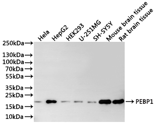
Western Blot
Positive WB detected in: Hela whole cell lysate(30µg), HepG2 whole cell lysate(30µg), HEK293 whole cell lysate(30µg), U-251MG whole cell lysate(30µg), SH-SY5Y whole cell lysate(30µg), Mouse brain tissue lysate(30µg), Rat brain tissue lysate(30µg)
All lanes: PEBP1 antibody at 1:1000
Secondary
Goat polyclonal to rabbit IgG at 1/40000 dilution
Predicted band size: 21 kDa
Observed band size: 21 kDa
Exposure time:1min
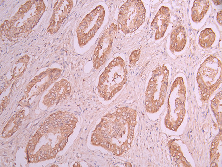
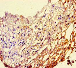
IHC image of CSB-RA903387A0HU diluted at 1:100 and staining in paraffin-embedded human prostate cancer performed on a Leica BondTM system. After dewaxing and hydration, antigen retrieval was mediated by high pressure in a citrate buffer (pH 6.0). Section was blocked with 10% normal goat serum 30min at RT. Then primary antibody (1% BSA) was incubated at 4°C overnight. The primary is detected by a Goat anti-rabbit polymer IgG labeled by HRP and visualized using 0.05% DAB.
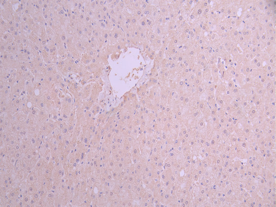
IHC image of CSB-RA903387A0HU diluted at 1:100 and staining in paraffin-embedded human liver tissue performed on a Leica BondTM system. After dewaxing and hydration, antigen retrieval was mediated by high pressure in a citrate buffer (pH 6.0). Section was blocked with 10% normal goat serum 30min at RT. Then primary antibody (1% BSA) was incubated at 4°C overnight. The primary is detected by a Goat anti-rabbit polymer IgG labeled by HRP and visualized using 0.05% DAB.
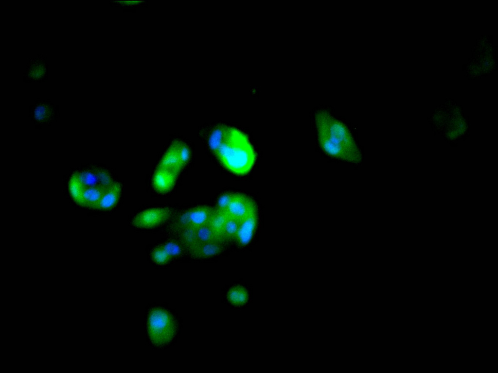
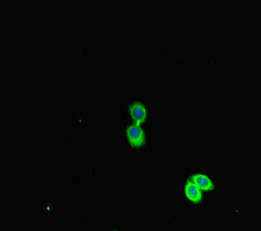
Immunofluorescence staining of HepG2 cell with CSB-RA903387A0HU at 1:50, counter-stained with DAPI. The cells were fixed in 4% formaldehyde, permeabilized using 0.2% Triton X-100 and blocked in 10% normal Goat Serum. The cells were then incubated with the antibody overnight at 4°C. The secondary antibody was Alexa Fluor 488-congugated AffiniPure Goat Anti-Rabbit IgG(H+L).
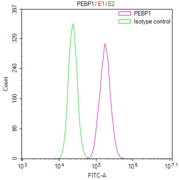
Overlay Peak curve showing PC-3 cells stained with CSB-RA903387A0HU (red line) at 1:100. The cells were fixed in 4% formaldehyde and permeated by 0.2% TritonX-100 for 10min. Then 10% normal goat serum to block non-specific protein-protein interactions followed by the antibody (1ug/1*106cells) for 45min at 4℃. The secondary antibody used was FITC-conjugated goat anti-rabbit IgG (H+L) at 1/200 dilution for 35min at 4℃.Control antibody (green line) was Rabbit IgG (1ug/1*106cells) used under the same conditions. Acquisition of >10,000 events was performed.
PEBP1 Antibody (CSB-PA017766HA01HU)
Validated Data
Immunohistochemistry of paraffin-embedded human thyroid tissue using CSB-PA017766HA01HU at dilution of 1:100
Immunofluorescent analysis of HepG2 cells using CSB-PA017766HA01HU at dilution of 1:100 and Alexa Fluor 488-congugated AffiniPure Goat Anti-Rabbit IgG(H+L)
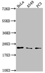
Western Blot
Positive WB detected in: Hela whole cell lysate, A549 whole cell lysate, PC3 whole cell lysate
All lanes: PEBP1 antibody at 1:2000
Secondary
Goat polyclonal to rabbit IgG at 1/50000 dilution
Predicted band size: 22 kDa
Observed band size: 22 kDa
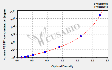
Human Phosphatidylethanolamine-binding protein 1(PEBP1) ELISA kit (CSB-EL017766HU)
Validated Data
Code: CSB-EL017766HU
Size: 96T,5×96T,10×96T
Sensitivity: 0.039 ng/mL
Detection Range: 0.156 ng/mL-10 ng/mL
These standard curves are provided for demonstration only. A standard curve should be generated for each set of samples assayed.
The following PEBP1 reagents supplied by CUSABIO are manufactured under a strict quality control system. Multiple applications have been validated and solid technical support is offered.
PEBP1 Antibodies for Homo sapiens (Human)
| Code | Product Name | Species Reactivity | Application |
|---|---|---|---|
| CSB-PA017766GA01HU | PEBP1 Antibody | Human,Mouse,Rat | ELISA,WB,IHC |
| CSB-PA948956 | PEBP1 Antibody | Human | ELISA,WB |
| CSB-PA004014 | PEBP1 Antibody | Human | WB, ELISA |
| CSB-PA614951 | PEBP1 Antibody | Human,Mouse,Rat | ELISA,WB,IHC |
| CSB-PA955522 | PEBP1 Antibody | Human,Mouse,Rat | ELISA,WB,IHC |
| CSB-PA017766HA01HU | PEBP1 Antibody | Human | ELISA, WB, IHC, IF |
| CSB-PA017766HB01HU | PEBP1 Antibody, HRP conjugated | Human | ELISA |
| CSB-PA017766HC01HU | PEBP1 Antibody, FITC conjugated | Human | |
| CSB-PA017766HD01HU | PEBP1 Antibody, Biotin conjugated | Human | ELISA |
| CSB-RA903387A0HU | PEBP1 Recombinant Monoclonal Antibody | Human, Mouse, Rat | ELISA, WB, IHC, IF, FC |
PEBP1 Proteins for Homo sapiens (Human)
| Code | Product Name | Source |
|---|---|---|
| CSB-YP017766HU CSB-EP017766HU CSB-BP017766HU CSB-MP017766HU CSB-EP017766HU-B |
Recombinant Human Phosphatidylethanolamine-binding protein 1 (PEBP1) | Yeast E.coli Baculovirus Mammalian cell In Vivo Biotinylation in E.coli |
PEBP1 ELISA Kit for Homo sapiens (Human)
| Code | Product Name | Sample Types | Sensitivity |
|---|---|---|---|
| CSB-EL017766HU | Human Phosphatidylethanolamine-binding protein 1(PEBP1) ELISA kit | serum, plasma, cell lysates | 0.039 ng/mL |

